You have no items in your shopping cart.
Cart summary
Item 1 of 6
Item 1 of 6
Goat anti-XRCC4-like factor / NHEJ1 Antibody
Catalog Number: orb19337

| Catalog Number | orb19337 |
|---|---|
| Category | Antibodies |
| Description | Goat polyclonal antibody to NHEJ1 |
| Species/Host | Goat |
| Clonality | Polyclonal |
| Tested applications | ELISA, FC, IF, IHC, WB |
| Reactivity | Canine, Human |
| Dilution range | ELISA: 1:64000, WB: 0.1-0.3 μg/ml, IHC-P: 2.5-3.8µg/ml |
| Conjugation | Unconjugated |
| MW | 33.3 |
| Target | XRCC4-like factor / NHEJ1 |
| Entrez | 79840 |
| Protein Sequence | QRPQLSKVKRKKPR |
| RRID | AB_10747536 |
| Storage | Aliquot and store at -20°C. Minimize freezing and thawing. |
| Buffer/Preservatives | Supplied at 0.5 mg/ml in Tris saline, 0.02% sodium azide, pH 7.3 with 0.5% bovine serum albumin. Aliquot and store at -20°C. Minimize freezing and thawing. |
| Alternative names | anti NHEJ1 antibody, anti XRCC4-like factor antibo Read more... |
| Note | For research use only |
| Application notes | ELISA: Peptide ELISA: antibody detection limit dilution 1:64000.WB: Approx 38kDa band observed in lysates of Human Skin, Testis and Thyroid gland (calculated size of 33.3kDa according to NP_079058.1). The observed molecular weight corresponds to findings with antibodies from other sources. The 38kDa band was successfully blocked by incubation with the immunizing peptide. Recommended concentration 0.1-0.3 μg/ml. |
| Expiration Date | 12 months from date of receipt. |

3.8 µg/ml staining of paraffin embedded Human Placenta. Steamed antigen retrieval with citrate buffer pH6, AP-staining.
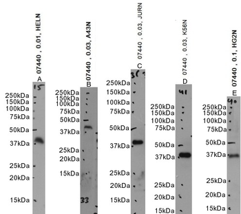
Primary incubation 1 hour at room temperature.Image A: HeLa nuclear cell lysate at primary Ab concentration 0.01 µg/ml, Images B, C, D: A431, Jurkat, K562 nuclear cell lysate at primary Ab concentration 0.03 µg/ml, Image E: HepG2 nuclear cell lysate at primary Ab concentration 0.1 µg/ml. (Loaded 35 µg protein in RIPA buffer, per lane). Detected by chemiluminescence.
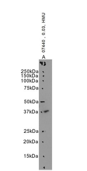
Primary incubation 1 hour at room temperature. Image A: Human Skeletal muscle lysate at primary Ab concentration 0.03 ug/ml. (Loaded 35 µg protein in RIPA buffer, per lane). Detected by chemiluminescence.
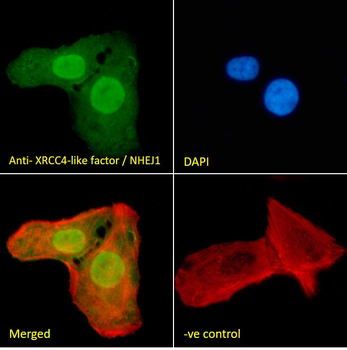
Immunofluorescence analysis of paraformaldehyde fixed U2OS cells, permeabilized with 0.15% Triton. Primary incubation 1hr (10 ug/ml) followed by Alexa Fluor 488 secondary antibody (2 ug/ml), showing strong nuclear staining. Actin filaments were stained with phalloidin (red) and the nuclear stain is DAPI (blue). Negative control: Unimmunized goat IgG (10 ug/ml) followed by Alexa Fluor 488 secondary antibody (2 ug/ml).
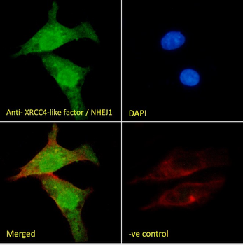
Immunofluorescence analysis of paraformaldehyde fixed HepG2 cells, permeabilized with 0.15% Triton. Primary incubation 1hr (10 ug/ml) followed by Alexa Fluor 488 secondary antibody (2 ug/ml), showing nuclear and cytoplasmic staining. Actin filaments were stained with phalloidin (red) and the nuclear stain is DAPI (blue). Negative control: Unimmunized goat IgG (10 ug/ml) followed by Alexa Fluor 488 secondary antibody (2 ug/ml).
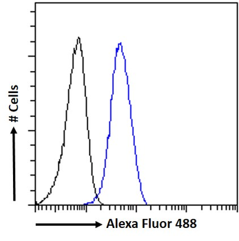
Flow cytometric analysis of paraformaldehyde fixed HepG2 cells (blue line), permeabilized with 0.5% Triton. Primary incubation 1hr (10 ug/ml) followed by Alexa Fluor 488 secondary antibody (1 ug/ml). IgG control: Unimmunized goat IgG (black line) followed by Alexa Fluor 488 secondary antibody.
