You have no items in your shopping cart.
Cart summary
Item 1 of 9
Item 1 of 9
Goat anti-NOS1 Antibody
Catalog Number: orb18337

| Catalog Number | orb18337 |
|---|---|
| Category | Antibodies |
| Description | Goat polyclonal antibody to NOS1 |
| Species/Host | Goat |
| Clonality | Polyclonal |
| Tested applications | ELISA, FC, IF, IHC |
| Reactivity | Bovine, Canine, Human, Mouse, Rat |
| Dilution range | ELISA: 1:16000, IHC-P: 2.5μg/ml, IF/ICC: 10μg/ml, FACS: 10ug/ml |
| Conjugation | Unconjugated |
| MW | 161; 164.6; 125 |
| Target | NOS1 |
| Entrez | 4842 |
| Protein Sequence | ESKKDTDEVFSS |
| RRID | AB_10754443 |
| Storage | Maintain refrigerated at 2-8°C for up to 2 weeks. For long term storage store at -20°C in small aliquots to prevent freeze-thaw cycles. |
| Buffer/Preservatives | Supplied at 0.5 mg/ml in Tris saline, 0.02% sodium azide, pH 7.3 with 0.5% bovine serum albumin. Aliquot and store at -20°C. Minimize freezing and thawing. |
| Alternative names | anti NOS1 antibody, anti nitric oxide synthase 1 ( Read more... |
| Note | For research use only |
| Application notes | ELISA: Peptide ELISA: antibody detection limit dilution 1:64000.IHC: This product has been sucessfully used for IHC on Olfactory bulb in mice (PMID: 20140458)WB: Approx 160-170kDa band observed in Human Skeletal Muscle and Mouse Brain lysates (calculated MW of 161kDa according to NP_000611.1). Recommended concentration: 0.3-1 μg/ml.Experiment Notes: Immunofluorescence: This product has been successfully used for IF as reported (PMID: 20140458). |
| Expiration Date | 12 months from date of receipt. |

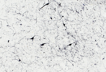
Immunohistochemical staining of mouse caudate-putamen using NOS1 antibody

Flow cytometric analysis of Kelly cells using NOS1 antibody

Immunofluorescence analysis of HeLa cells using NOS1 antibody

Immunohistochemical staining of Human Cortex using NOS1 antibody
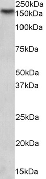
1 µg/mL staining of Mouse Brain lysate (35 µg protein in RIPA buffer). Detected by chemiluminescence.
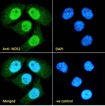
Immunofluorescence analysis of paraformaldehyde fixed HeLa cells, permeabilized with 0.15% Triton. Primary incubation 1 hr (10 µg/mL) followed by Alexa Fluor 488 secondary antibody (2 µg/mL), showing nuclear staining. The nuclear stain is DAPI (blue). Negative control: Unimmunized goat IgG (10 µg/mL) followed by Alexa Fluor 488 secondary antibody (2 µg/mL).
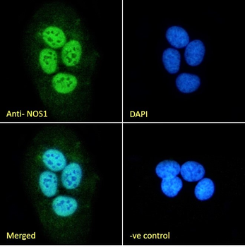
Immunofluorescence analysis of paraformaldehyde fixed U2OS cells, permeabilized with 0.15% Triton. Primary incubation 1 hr (10 µg/mL) followed by Alexa Fluor 488 secondary antibody (2 µg/mL), showing nuclear staining. The nuclear stain is DAPI (blue). Negative control: Unimmunized goat IgG (10 µg/mL) followed by Alexa Fluor 488 secondary antibody (2 µg/mL).
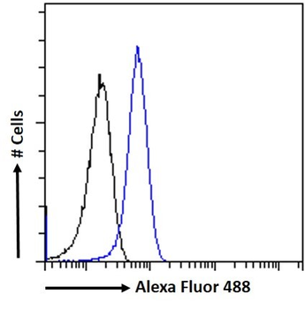
Flow cytometric analysis of paraformaldehyde fixed HeLa cells (blue line), permeabilized with 0.5% Triton. Primary incubation 1 hr (10 µg/mL) followed by Alexa Fluor 488 secondary antibody (1 µg/mL). IgG control: Unimmunized goat IgG (black line) followed by Alexa Fluor 488 secondary antibody.
Immunostaining of 30 µm thick cryosections of PFA-perfused Human Hypothalamus, antigen retrieval with citrate buffer Ph 6 at 80C for 30 min, HRP-staining with Ni-DAB after Biotin-SP-antigoat amplification.
- Item 1 of 1

