You have no items in your shopping cart.
Cart summary
Item 1 of 6
Item 1 of 6
Goat anti-CD32 / FCGR2B Antibody
Catalog Number: orb19010

| Catalog Number | orb19010 |
|---|---|
| Category | Antibodies |
| Description | Goat polyclonal antibody to CD32b |
| Species/Host | Goat |
| Clonality | Polyclonal |
| Tested applications | ELISA, FC, IF, IHC, WB |
| Reactivity | Human |
| Dilution range | ELISA: 1:32000, WB: 0.3-1 μg/ml |
| Conjugation | Unconjugated |
| MW | 34.0; 31.9; 31.9; 34.0; 33.4 |
| Target | CD32 / FCGR2B |
| Entrez | 2213 |
| Protein Sequence | PDALEEPDDQNRI |
| RRID | AB_10749756 |
| Storage | Maintain refrigerated at 2-8°C for up to 2 weeks. For long term storage store at -20°C in small aliquots to prevent freeze-thaw cycles. |
| Buffer/Preservatives | Supplied at 0.5 mg/ml in Tris saline, 0.02% sodium azide, pH 7.3 with 0.5% bovine serum albumin. Aliquot and store at -20°C. Minimize freezing and thawing. |
| Alternative names | anti-CD32 antibody, anti- CD32 antigen antibody, a Read more... |
| Note | For research use only |
| Application notes | ELISA: Peptide ELISA: antibody detection limit dilution 1:16000.WB: Approx 34kDa band observed in Human myeloid erythroblast cell line K562 and Human monocytic cell line U937 lysates (calculated MW of 34.0kDa according to NP_003992.3;). In transfected HEK293 transiently expressing CD32 a band of approx. 40kDa (glycoprotein) is observed. This band is not observed in the non-transfected HEK293. Recommended concentration: 0.3-1 μg/ml. |
| Expiration Date | 12 months from date of receipt. |

1 μg/ml staining of Daudi cell lysate (RIPA buffer, 35 μg protein in RIPA buffer). Detected by chemiluminescence.
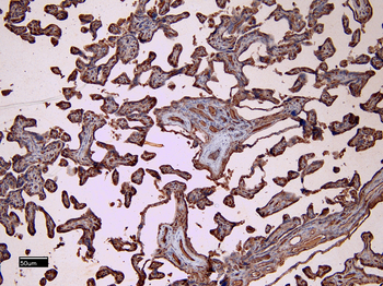
8 μg/ml staining of paraffin embedded Human Placenta. Heat induced antigen retrieval with citrate buffer pH 6, HRP-staining.
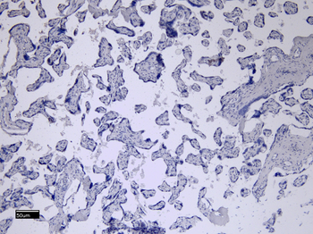
Negative Control showing staining of paraffin embedded Human Placenta, with no primary antibody.
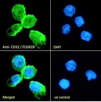
Immunofluorescence analysis of paraformaldehyde fixed THP-1 cells immobilized on coverslip, permeabilized with 0.15% Triton. Primary incubation 1hr (10 μg/ml) followed by Alexa Fluor 488 secondary antibody (2 μg/ml), showing membrane staining. The nuclear stain is DAPI (blue). Negative control: Unimmunized goat IgG (10 μg/ml) followed by Alexa Fluor 488 secondary antibody (2 μg/ml).
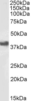
0.5 μg/ml staining of Human Placenta lysate (RIPA buffer, 35 μg protein in RIPA buffer). Detected by chemiluminescence.

Flow cytometric analysis of paraformaldehyde fixed K562 cells (blue line), permeabilized with 0.5% Triton. Primary incubation 1hr (10 μg/ml) followed by Alexa Fluor 488 secondary antibody (1 μg/ml). IgG control: Unimmunized goat IgG (black line) followed by Alexa Fluor 488 secondary antibody.
