You have no items in your shopping cart.
Cart summary
Item 1 of 5
Item 1 of 5
Glypican 3 Antibody / GPC3
Catalog Number: orb749525

| Catalog Number | orb749525 |
|---|---|
| Category | Antibodies |
| Description | Glypican-3 (GPC3) is a glycosylphospatidyl inositol-anchored membrane protein, which may also be found in a secreted form. Anti-GPC3 has been identified as a useful tumor marker for the diagnosis of hepatocellular carcinoma (HCC), hepatoblastoma, melanoma, testicular germ cell tumors, and Wilm's tumor. In patients with HCC, GPC3 is overexpressed in neoplastic liver tissue and elevated in serum, but is undetectable in normal liver, benign liver, and the serum of healthy donors. GPC3 expression is also found to be higher in HCC liver tissue than in cirrhotic liver or liver with focal lesions such as dysplastic nodules and areas of hepatic adenoma (HA) with malignant transformation. In the context of testicular germ cell tumors, GPC3 expression is up regulated in certain histologic subtypes, specifically yolk sac tumors and choriocarcinoma. A high level of GPC3 expression is also found in some types of embryonal tumors, such as Wilm�s tumor and hepatoblastoma, with a low or undetectable expression in normal adjacent tissue. In patients with thyroid cancer, expression of GPC3 is dramatically enhanced in certain types of cancers: 100% in follicular carcinoma and 70% in papillary carcinoma. Expression of GPC3 in follicular carcinoma is significantly higher than that of follicular adenoma. In contrast, GPC3 is not expressed in anaplastic carcinoma. |
| Species/Host | Mouse |
| Clonality | Monoclonal |
| Clone Number | 1G12 |
| Tested applications | FACS, IF, IHC-P, WB |
| Reactivity | Human |
| Isotype | Mouse IgG1, kappa |
| Immunogen | A recombinant fragment containing amino acids 511-580 from the human protein was used as the immunogen for the Glypican-3 antibody. |
| Dilution range | Western blot: 1-2ug/ml,Flow cytometry: 1-2ug/million cells,Immunofluorescence: 1-2ug/ml,Immunohistochemistry (FFPE): 0.5-1ug/ml for 30 min at RT |
| Purity | Protein G affinity chromatography |
| Conjugation | Unconjugated |
| Formula | 0.2 mg/ml in 1X PBS with 0.1 mg/ml BSA (US sourced) and 0.05% sodium azide |
| Hazard Information | This Glypican-3 antibody is available for research use only. |
| UniProt ID | P51654 |
| Storage | Store the Glypican-3 antibody at 2-8°C (with azide) or aliquot and store at -20°C or colder (without azide). |
| Buffer/Preservatives | 0.2 mg/ml in 1X PBS with 0.1 mg/ml rAlbumin (US sourced) and 0.05% sodium azide |
| Note | For research use only |
| Application notes | Optimal dilution of the Glypican-3 antibody should be determined by the researcher.1. Staining of formalin-fixed tissues requires boiling tissue sections in 10mM Tris with 1mM EDTA, pH 9.0, for 10-20 min followed by cooling at RT for 20 min2. The prediluted format is supplied in a dropper bottle and is optimized for use in IHC. After epitope retrieval step (if required), drip mAb solution onto the tissue section and incubate at RT for 30 min. |
| Expiration Date | 12 months from date of receipt. |
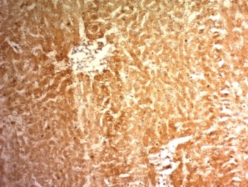
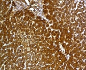
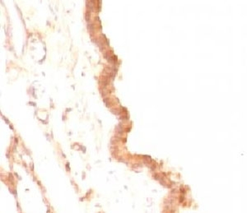
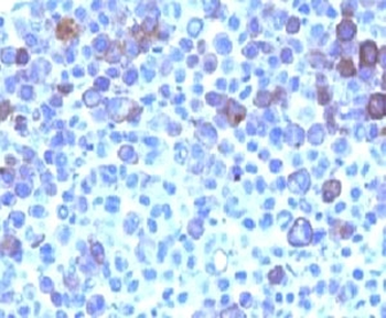
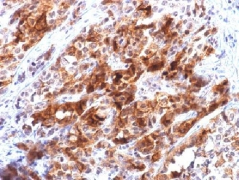
IHC: Formalin-fixed, paraffin-embedded human hepatocellular carcinoma stained with Glypican-3 antibody (clone 1G12).

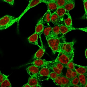
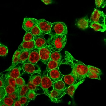
Immunofluorescent staining of methanol-fixed HepG2 cells with Glypican-3 antibody (green, clone 1G12) and Reddot nuclear stain (red).
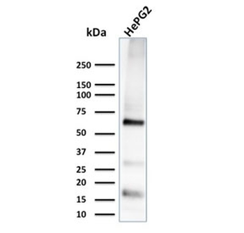
Western blot testing of human HePG2 cell lysate with Glypican-3 antibody (clone 1G12). Expected molecular weight 66-115 kDa depending on glycosylation level.
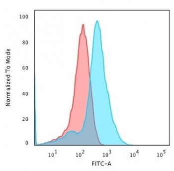
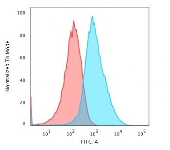
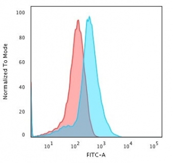
Flow cytometry testing of methanol-fixed human HepG2 cells with Glypican-3 antibody (clone 1G12); Red = isotype control, Blue = Glypican-3 antibody.

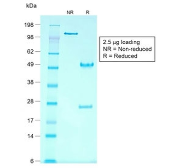
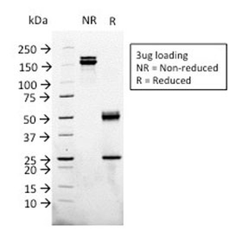
SDS-PAGE analysis of purified, BSA-free Glypican-3 antibody (clone 1G12) as confirmation of integrity and purity.
- Item 1 of 6
- Item 1 of 6
- Item 1 of 5
- Item 1 of 5
- Item 1 of 5