You have no items in your shopping cart.
Cart summary
Item 1 of 6
Item 1 of 6
GLS2 Antibody
Catalog Number: orb1239416

| Catalog Number | orb1239416 |
|---|---|
| Category | Antibodies |
| Description | GLS2 Antibody |
| Species/Host | Rabbit |
| Clonality | Polyclonal |
| Tested applications | ELISA, IF, IHC-P, WB |
| Reactivity | Human, Mouse, Rat |
| Isotype | IgG |
| Immunogen | GLS2 antibody was raised against an 18 amino acid synthetic peptide near the center terminus of human GLS2.The immunogen is located within amino acids 300 - 350 of GLS2. |
| Antibody Type | Primary Antibody |
| Concentration | 1 mg/mL |
| Form/Appearance | Liquid |
| Conjugation | Unconjugated |
| MW | Predicted: 36, 55, 62, 66 kDa Observed: 55, 62 kDa |
| Target | GLS2 |
| UniProt ID | Q9UI32 |
| NCBI | NP_037399 |
| Storage | Maintain refrigerated at 2-8°C for up to 2 weeks. For long term storage store at -20°C in small aliquots to prevent freeze-thaw cycles. |
| Buffer/Preservatives | GLS2 Antibody is supplied in PBS containing 0.02% sodium azide. |
| Alternative names | GLS2 Antibody: GA, GLS, LGA, hLGA, GA, Glutaminase Read more... |
| Note | For research use only |
| Application notes | GLS2 antibody can be used for detection of GLS2 by Western blot at 0.5 - 1 μg/mL. Antibody can also be used for immunohistochemistry starting at 5 μg/mL. For immunofluorescence start at 20 μg/mL.Antibody validated: Western Blot in rat samples; Immunohistochemistry in mouse and rat samples and Immunofluorescence in mouse and rat samples. All other applications and species not yet tested. |
| Expiration Date | 12 months from date of receipt. |

Immunofluorescence Validation of GLS2 in Mouse Brain. Immunofluorescent analysis of 4% paraformaldehyde-fixed mouse brain labeling GLS2 with orb1239416 at 20 µg g/mL, followed by goat anti-rabbit IgG secondary antibody at 1/500 dilution (green) and DAPI antibody (blue).
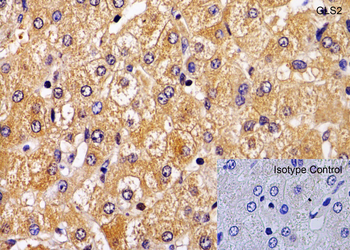
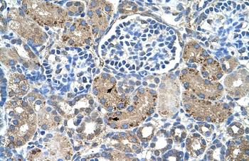
Immunohistochemistry Validation of GLS2 in Human Liver. Immunohistochemical analysis of paraffin-embedded human liver tissue using anti-GLS2 antibody (orb1239416) at 1 µg/mL. Tissue was fixed with formaldehyde and blocked with 10% serum for 1 h at RT; antigen retrieval was by heat mediation with a citrate buffer (pH6). Samples were incubated with primary antibody overnight at 4°C. A goat anti-rabbit IgG H&L (HRP) at 1/250 was used as secondary. Counter stained with Hematoxylin.
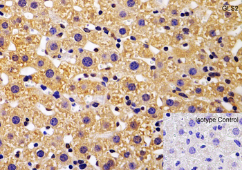
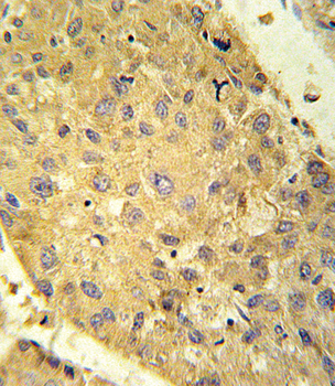
Immunohistochemistry Validation of GLS2 in Mouse Liver. Immunohistochemical analysis of paraffin-embedded mouse liver tissue using anti-GLS2 antibody (orb1239416) at 1 µg/mL. Tissue was fixed with formaldehyde and blocked with 10% serum for 1 h at RT; antigen retrieval was by heat mediation with a citrate buffer (pH6). Samples were incubated with primary antibody overnight at 4°C. A goat anti-rabbit IgG H&L (HRP) at 1/250 was used as secondary. Counter stained with Hematoxylin.
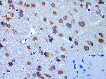
Immunohistochemistry Validation of GLS2 in Rat Brain. Immunohistochemical analysis of paraffin-embedded rat brain tissue using anti-GLS2 antibody (orb1239416) at 2 µg/mL. Tissue was fixed with formaldehyde and blocked with 10% serum for 1 h at RT; antigen retrieval was by heat mediation with a citrate buffer (pH6). Samples were incubated with primary antibody overnight at 4°C. A goat anti-rabbit IgG H&L (HRP) at 1/250 was used as secondary. Counter stained with Hematoxylin.

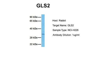
WB Validation in Human Pancreas. Loading: 10 µg of lysate Antibodies: GLS2, orb1239416, 2 µg/mL, 1 h incubation at RT in 5% NFDM/TBST. Secondary: Goat Anti-Rabbit IgG HRP conjugate at 1:10000 dilution.
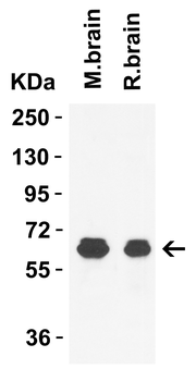
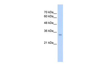
WB Validation in Mouse and Rat Brain. Loading: 15 µg of lysate Antibodies: GLS2, orb1239416, 1 µg/mL, 1 h incubation at RT in 5% NFDM/TBST. Secondary: Goat Anti-Rabbit IgG HRP conjugate at 1:10000 dilution.
- Item 1 of 3
GLS2 Rabbit Polyclonal Antibody [orb578865]
IHC, WB
Bovine, Canine, Equine, Guinea pig, Mouse, Porcine, Rabbit, Rat, Zebrafish
Human
Rabbit
Polyclonal
Unconjugated
100 μl - Item 1 of 3
GLS2 Antibody (C-term E513) [orb1788150]
IHC-P, WB
Human, Mouse, Rat
Rabbit
Polyclonal
Unconjugated
100 μl - Item 1 of 2
- Item 1 of 2
- Item 1 of 2