You have no items in your shopping cart.
Cart summary
Item 1 of 3
Item 1 of 3
Gli-2 antibody
Catalog Number: orb345479

| Catalog Number | orb345479 |
|---|---|
| Category | Antibodies |
| Description | Gli-2 antibody |
| Species/Host | Rabbit |
| Clonality | Polyclonal |
| Tested applications | ELISA, IHC, WB |
| Reactivity | Mouse |
| Isotype | IgG |
| Immunogen | This affinity purified antibody was prepared from whole rabbit serum produced by repeated immunizations with a synthetic peptide corresponding to amino acids from an internal region of Mouse Gli-2. |
| Concentration | 1.02 mg/mL |
| Dilution range | ELISA: 1:15,000 - 1:60,000, IHC: 2 µg/ml to 20 µg/ml, WB: 1:500 - 1:2,000 |
| Form/Appearance | Liquid (sterile filtered) |
| Purity | This affinity purified antibody is directed against mouse Gli-2 protein. The product was affinity purified from monospecific antiserum by immunoaffinity chromatography. A BLAST analysis was used to suggest cross-reactivity with Gli-2 from mouse and rat sources based on 100% sequence homology with the immunogen. Reactivity with Gli-2 from other sources is not known. |
| Conjugation | Unconjugated |
| UniProt ID | Q8K0K3 |
| NCBI | 124487481 |
| Storage | Store vial at -20° C prior to opening. Aliquot contents and freeze at -20° C or below for extended storage. Avoid cycles of freezing and thawing. Centrifuge product if not completely clear after standing at room temperature. This product is stable for several weeks at 4° C as an undiluted liquid. Dilute only prior to immediate use. |
| Buffer/Preservatives | 0.01% (w/v) Sodium Azide |
| Alternative names | rabbit anti-Gli-2 antibody, Gli 2, Gli2, zinc fing Read more... |
| Note | For research use only |
| Application notes | This antibody has been tested for use in ELISA, immunohistochemistry and western blot. Specific conditions for reactivity should be optimized by the end user. See figure legend for expectations by WB and IHC. Multiple splice variants have been reported for this protein. |
| Expiration Date | 12 months from date of receipt. |
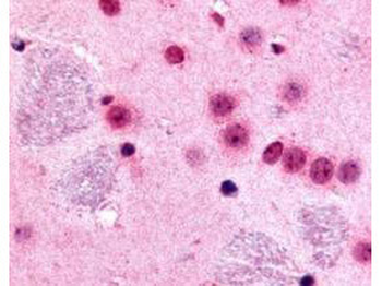
Biorbyt's Affinity Purified anti-mouse Gli-2 antibody was used at 10 µg/ml to evaluate staining on several mouse tissues. Moderate to strong staining was seen on many tissues with low background staining. This image shows Gli-2 staining of mouse brain. Tissue was formalin-fixed and paraffin embedded.
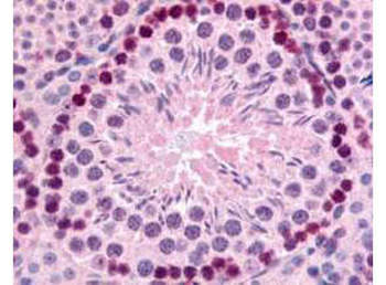
Biorbyt's Affinity Purified anti-mouse Gli-2 antibody was used at 10 µg/ml to evaluate staining on several mouse tissues. Moderate to strong staining was seen on many tissues, with low background staining. This image shows Gli-2 staining of mouse testis. Tissue was formalin-fixed and paraffin embedded.
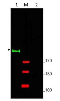
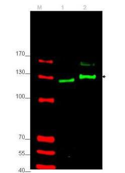
Western blot using Biorbyt's Affinity Purified anti-Gli-2 antibody shows detection of a predominant band at ~190 kDa corresponding to Gli-2 (arrowhead) in mouse brain whole cell lysate (p/n orb348715) (lane 1). Pre-incubation of antibody with immunizing peptide completely blocks staining of this band (lane 2). Load 25 µg of lysate was resolved on a 4-8% Tris-glycine gel by SDS-PAGE and transferred onto nitrocellulose. After blocking with 5% goat serum and 0.5% BLOTTO in PBS, the membrane was probed with the primary antibody diluted to 1:750. Incubation was at room temperature for 2 h followed by washes and reaction with a 1:10000 dilution of IRDye® 800 conjugated Gt-a-Rabbit IgG (H&L) MX10 for 45 min at room temperature. Molecular weight markers are shown (M) using the 700 nm channel (red).
- Item 1 of 3
- Item 1 of 3
- Item 1 of 2
- Item 1 of 1
Gli2 antibody [orb157157]
FC, ICC, IHC-P
Bovine, Canine, Equine, Porcine, Rabbit
Human, Mouse, Rat
Rabbit
Polyclonal
Unconjugated
200 μl, 50 μl, 100 μl 


Submit a review
Filter by Rating

- 5 stars

- 4 stars
- 3 stars
- 2 stars
- 1 stars
