You have no items in your shopping cart.
Cart summary
Item 1 of 6
Item 1 of 6

| Catalog Number | orb345367 |
|---|---|
| Category | Antibodies |
| Description | GFP antibody |
| Species/Host | Rabbit |
| Clonality | Polyclonal |
| Tested applications | ELISA, FC, IF, IHC, IP, WB |
| Reactivity | Other |
| Isotype | IgG |
| Immunogen | The immunogen is a Green Fluorescent Protein (GFP) fusion protein corresponding to the full length amino acid sequence (246aa) derived from the jellyfish Aequorea victoria. |
| Concentration | 1.25 mg/mL |
| Dilution range | ELISA: 1:20,000 - 1:120,000, FC: User Optimized, IHC: 1:200 - 1:3,000, IF: 1:500 - 1:5,000, IP: User Optimized, WB: 1:500 - 1:5,000 |
| Form/Appearance | Liquid (sterile filtered) |
| Purity | Anti-GFP antibody was prepared from monospecific antiserum by immunoaffinity chromatography using Green Fluorescent Protein (Aequorea victoria) coupled to agarose beads followed by solid phase adsorption(s) to remove any unwanted reactivities. Assay by immunoelectrophoresis resulted in a single precipitin arc against anti-Rabbit Serum and purified and partially purified Green Fluorescent Protein (Aequorea victoria). No reaction was observed against Human, Mouse or Rat serum proteins. |
| Conjugation | Unconjugated |
| UniProt ID | P42212 |
| Storage | Store Anti-GFP Antibody at -20° C prior to opening. Aliquot contents and freeze at -20° C or below for extended storage. Avoid cycles of freezing and thawing. Centrifuge product if not completely clear after standing at room temperature. GFP antibody is stable for several weeks at 4° C as an undiluted liquid. Dilute only prior to immediate use. |
| Buffer/Preservatives | 0.01% (w/v) Sodium Azide |
| Alternative names | rabbit anti-GFP antibody, Green Fluorescent Protei Read more... |
| Note | For research use only |
| Application notes | Anti-GFP antibody is designed to detect GFP and its variants. GFP antibody has been tested by western blot and ELISA. This product can be used to detect GFP by ELISA (sandwich or capture) for the direct binding of antigen and recognizes wild type, recombinant and enhanced forms of GFP. Biotin conjugated polyclonal anti-GFP used in a sandwich ELISA is well suited to titrate GFP in solution using this antibody in combination with Rockland's monoclonal anti-GFP (600-301-215) using either form of the antibody as the capture or detection antibodies. However, use the monoclonal form only for the detection of wild type or recombinant GFP as this form does not sufficiently detect 'enhanced' GFP. The detection antibody is typically conjugated to biotin and subsequently reacted with streptavidin conjugated HRP (code # S000-03). Fluorochrome conjugated polyclonal anti-GFP can be used to detect GFP by immunofluorescence microscopy in prokaryotic (E.coli) and eukaryotic (CHO cells) expression systems and can detect GFP containing inserts. Significant amplification of signal is achieved using fluorochrome conjugated polyclonal anti-GFP relative to the fluorescence of GFP alone. For immunoblotting use either alkaline phosphatase or peroxidase conjugated polyclonal anti-GFP to detect GFP or GFP containing proteins on western blots. Optimal titers for applications should be determined by the researcher. |
| Expiration Date | 12 months from date of receipt. |
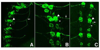
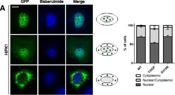
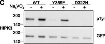
Widowati, Esti Wahyu et al. Functional characterization of DYRK1A missense variants associated with a syndromic form of intellectual deficiency and autism Biol Open, 7, (2018)

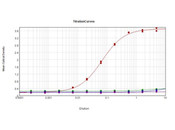
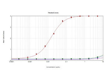
ELISA results of purified Rabbit Anti-GFP Antibody. Each well was coated in 10 µg of antigen GFP [Red Line], human IgG [Green Line], Mouse IgG [Blue Line], and Rat IgG [Purple Line]. The starting dilution of antibody was 5 µg/mL and the X-axis represents the Log10 of a 3-fold dilution. The titer is 1:67700. This titration is a 4-parameter curve fit where the IC50 is defined as the titer of the antibody. Assay performed using 1% Fish Gel, TMB Substrate, and Goat Anti-Rabbit IgG Antibody HRP (p/n orb347654).

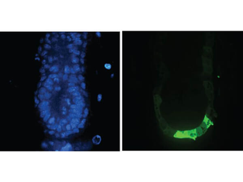
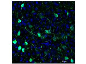
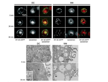
Immuno-microscopy of Rabbit anti-GFP antibody. Monocyte derived dendritic cells and dermal macrophages were challenged and directly visualized with eGFP labeled Dengue virus to localize sequestration of virus particles in the different cells (upper). The location of the GFP was confirmed by TEM (lower magnified view) using Biorbyt rabbit anti GFP Primary antibody (1:200) and a gold labeled secondary antibody.
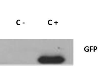
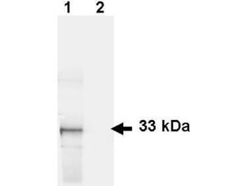
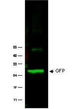
Western Blot of Rabbit anti-GFP antibody. Lane 1: 293FT cells transfected with CDK4 dominant negative (C-). Lane 2: 293FT cells poitive control (C+). Load: 25 µg per lane. Primary antibody: GFP antibody at 1:400 for overnight at 4°C. Secondary antibody: IRDye800™ rabbit secondary antibody at 1:10000 for 45 min at RT. Block: 5% BLOTTO overnight at 4°C. Predicted/Observed size: 27 kDa for GFP.
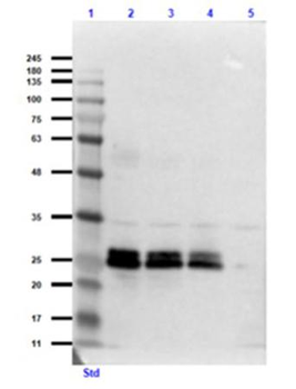
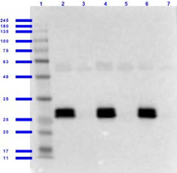
Western Blot of Rabbit Anti-GFP Antibody. Lane 1: Opal Prestained Molecular Weight Ladder. Lane 2: GFP (p/n orb345957) / HeLa Lysate (p/n orb348668) [0.1 µg/10.0 µg]. Lane 3: GFP (p/n orb345957) / HeLa Lysate (p/n orb348668) [0.05 µg/10.0 µg]. Lane 4: GFP (p/n orb345957) / HeLa Lysate (p/n orb348668) [0.03 µg/10.0 µg]. Lane 5: HeLa Lysate (p/n orb348668) [10.0 µg]. Primary Antibody: Rabbit Anti-GFP Antibody at 1.0 µg/mL overnight at 2-8°C. Secondary Antibody: Goat Anti-Rabbit IgG MX 10 Peroxidase (p/n orb347654) at 1:70000 for 30 mins at RT. Block: Blocking Buffer for Fluorescent Western Blotting (p/n orb348637) for 60 mins at RT. Expect: ~27 kDa.

Western Blot of Rabbit anti-GFP antibody. Lane 1: Wild type GFP (0.1 µg) was used to spike HeLa whole cell lysate. Lane 2: none. Load: 30 µg per lane. Primary antibody: GFP antibody at 1:1000 for overnight at 4°C. Secondary antibody: IRDye800™ Goat-a-Rabbit IgG [H&L] MX10 at 1:10000 for 45 min at RT. Block: 5% BLOTTO in PBS overnight at 4°C. Predicted/Observed size: 27 kDa for epitope tag GFP. Other band(s): none.

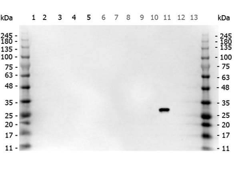
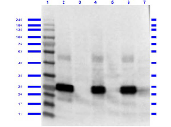
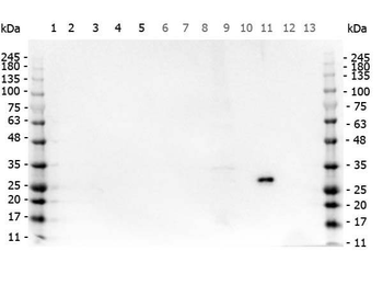
Western Blot of Rabbit anti-GFP antibody. Marker: Opal Pre-stained ladder. Lane 1: HEK293 lysate (p/n orb348669). Lane 2: HeLa Lysate (p/n orb348668). Lane 3: CHO/K1 Lysate. Lane 4: MDA-MB-231 (p/n orb348700). Lane 5: A431 Lysate (p/n orb348665). Lane 6: Jurkat Lysate (p/n orb348674). Lane 7: NIH/3T3 Lysate (p/n orb348714). Lane 8: E-coli HCP Control (p/n orb342262). Lane 9: FLAG Positive Control Lysate (p/n orb348661). Lane 10: Red Fluorescent Protein (p/n orb345960). Lane 11: Green Fluorescent Protein (p/n orb345957). Lane 12: Glutathione-S-Transferase Protein (p/n orb345956). Lane 13: Maltose Binding Protein. Load: 10 µg of lysate or 50 ng of purified protein per lane. Primary antibody: GFP antibody at 1 ug/ml overnight at 4C. Secondary antibody: Peroxidase rabbit secondary antibody (p/n orb347654) at 1:30000 for 60 min at RT. Blocking Buffer: 1% Casein-TTBS for 30 min at RT. Predicted/Observed size: 30 kDa for GFP.
- Item 1 of 28
- Item 1 of 28
- Item 1 of 28
- Item 1 of 6
- Item 1 of 6


Submit a review
Filter by Rating

- 5 stars

- 4 stars
- 3 stars
- 2 stars
- 1 stars
