You have no items in your shopping cart.
Cart summary
Item 1 of 6
Item 1 of 6
GFP antibody
Catalog Number: orb345330

| Catalog Number | orb345330 |
|---|---|
| Category | Antibodies |
| Description | GFP antibody |
| Species/Host | Mouse |
| Clonality | Monoclonal |
| Clone Number | 9F9.F9 |
| Tested applications | DOT, ELISA, FC, IF, IHC, IP, WB |
| Reactivity | Other |
| Isotype | IgG |
| Immunogen | Recombinant Green Fluorescent Protein (GFP) fusion protein corresponding to the full length amino acid sequence (246 aa) derived from the jellyfish Aequorea victoria. |
| Concentration | 1.0 mg/mL |
| Dilution range | ELISA: 1:80,000 - 1:500,000, FC: User Optimized, IHC: 1:1,000 - 1:5,000, IF: User Optimized, WB: 1:3000 - 1:30,000 |
| Form/Appearance | Liquid (sterile filtered) |
| Purity | GFP Monoclonal Antibody was prepared from tissue culture supernatant by Protein A affinity chromatography. Assay by Immunoelectrophoresis resulted in a single precipitin arc against anti-Mouse Serum. Reactivity is observed against recombinant Green Fluorescent Protein (000-001-215) from Aequorea victoria by both Western blot and ELISA. No reaction is seen against RFP. |
| Conjugation | Unconjugated |
| UniProt ID | P42212 |
| Storage | Store mouse anti-GFP at -20° C prior to opening. Aliquot contents and freeze at -20° C or below for extended storage. Avoid cycles of freezing and thawing. Centrifuge product if not completely clear after standing at room temperature. This product is stable for several weeks at 4° C as an undiluted liquid. Dilute only prior to immediate use. |
| Buffer/Preservatives | 0.01% (w/v) Sodium Azide |
| Alternative names | mouse anti-GFP antibody, Green Fluorescent Protein Read more... |
| Note | For research use only |
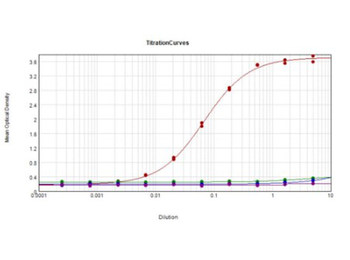
| Application notes | Monoclonal anti-GFP is designed to detect enhanced GFP and GFP containing recombinant proteins. Tested in ELISA, IP, and WB and suitable in FACS, IHC, IF. This antibody can be used to detect GFP by ELISA (sandwich or capture) for the direct binding of antigen. Biotin conjugated monoclonal anti-GFP is well suited to titrate GFP in a sandwich ELISA in combination with Rockland's polyclonal anti-GFP (600-101-215) as the capture antibody. Only use the monoclonal form for the detection of enhanced or recombinant GFP. Polyclonal anti-GFP detects all variants of GFP tested to date. The biotin conjugated detection antibody is typically used with streptavidin conjugated HRP (code # S000-03) or other streptavidin conjugates. The use of polyclonal anti-GFP results in significant amplification of signal when fluorochrome conjugated polyclonal anti-GFP is used relative to the fluorescence of GFP alone. For immunoblotting use either alkaline phosphatase or peroxidase conjugated anti-GFP to detect GFP or GFP containing proteins on western blots. Optimal titers for applications should be determined by the researcher. |
| Expiration Date | 12 months from date of receipt. |
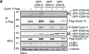
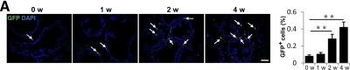
Active Cyclin Y/CDK16 complexes induce autophagy.a NIH3T3 cells stably expressing mCherry-GFP-LC3 were transfected with HA-CDK16 and Cyclin Y-Flag as indicated or treated for 2 h with EBSS or for 4 h with 200 nM Bafilomycin A1 (Baf. A1) and lysates were immunoblotted as indicated. KR kinase-deficient CDK16 mutant, AA CDK16 binding deficient Cyclin Y mutant (n=3). b Representative confocal images of the NIH3T3-mCherry-GFP-LC3 cells treated as in panel a. Staining of the HA-CDK16 in purple identified transfected cells. Autophagosomes (yellow dots) and autolysosomes (red dots) were detected by an overlay of the GFP and mCherry fluorescent signals. Scale bar: 20 µM. c Quantification of autophagosomes (yellow dots) and autolysosomes (red dots) of cells shown in panel b. Statistical significance was measured via unpaired and two-tailed Student's t-tests and is presented as follows: **p < 0.01. All error bars indicate SD (n=3; 50 cells counted for each replicate; wt vs. KR: t = 5.707, df = 4; wt vs. AA: t = 5.557, df = 4). d GFP-tagged CDK14, CDK15 or CDK16 were expressed with or without Cyclin Y-Flag in HeLa cells. Cyclin Y was immunoprecipitated with a Flag antibody (IP). Lysates were immunoblotted with the indicated antibodies (WCL). (n=4) e Representative confocal images of HeLa cells treated as in panel d. Fluorescent GFP signals identified CDK expressing cells. Endogenous LC3 (red) was used to measure autophagy with the 4E12 antibody. Scale bar: 50 µM. f Quantification of the LC3 dots shown in panel e. Statistical significance was measured via unpaired and two-tailed Student's t-tests and is presented as follows: ****p < 0.0001. All error bars indicate SD. (n=1; 100 cells were counted for each treatment; control vs. Cyclin Y/CDK16: t = 6.771, df = 14; Cyclin Y/CDK14 vs. Cyclin Y/CDK16: t = 6.855, df = 14; Cyclin Y/CDK15 vs. Cyclin Y/CDK16: t = 7.139, df = 15). n biological independent replicate. SD standard deviation.
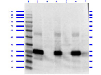
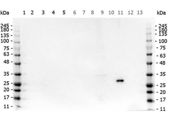
Immunoprecipitation/Western Blot using GFP Protein. Lane 1: Opal Prestained Molecular Weight Marker. Lane 2: GFP Input (p/n orb345957) Reduced [10 µL]. Primary IP Antibody: Mouse Anti-GFP (p/n orb345329) at 10 µg overnight at 2-8°C. Secondary Antibody: TrueBlot Anti-Mouse Ig IP Agarose Beads at 500 µg for 1 hr at RT. Buffer: BlockOut Buffer (p/n orb348644) for 30 mins at RT. Exposure: 7 sec.
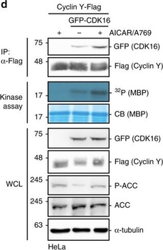
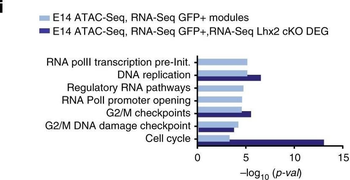
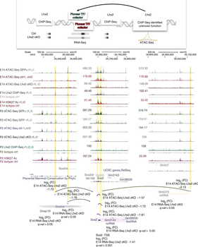
Protein microarray screen for the identification of AMPK substrates.a Schematic representation of the ProtoArray based screen with approximately 9000 human proteins using AMPK. b Details of two sub-arrays incubated with or without AMPK with marked substrates are shown. c GST-CDK16, Cyclin Y-His6 and GST were incubated in the presence of [γ-32P]-ATP with AMPK. Phosphorylation was determined by autoradiography (32P, top). Proteins were visualized by Coomassie blue staining (CB, bottom; n=2). d HeLa cells were transfected with vectors expressing GFP-CDK16 and Cyclin Y-Flag and treated for 1 h with 0.5 mM AICAR/50 µM A769662 (A769) as indicated. Cyclin Y-Flag was immunoprecipitated with Flag antibodies (IP) and immunoblotted against CDK16 and Cyclin Y or used for in vitro kinase assays with myeloid basic protein (MBP) as substrate. Autoradiographs (32P) and Coomassie blue staining (CB) of MBP are displayed. Whole cell lysates (WCL) were immunoblotted with the indicated antibodies (n=3). e Quantification of CDK16 co-immunoprecipitated with Cyclin Y. Statistical significance was measured via unpaired and two-tailed Student's t-tests and is presented as follows: **p < 0.01, and ***p < 0.001. All error bars indicate SD (n=3; Cyclin Y + AICAR/A769 vs. Cyclin Y/CDK16: t = 8.719, df = 4; Cyclin Y/CDK16 vs. Cyclin Y/CDK16 + AICAR/A769: t = 5.595, df = 4). n biological independent replicate. SD standard deviation.
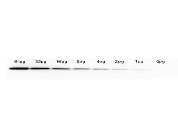
Western Blot of anti-GFP monoclonal antibody. Lane 1: 64 pg of recombinant GFP protein (p/n orb345957) were spiked into a HeLa cell-derived lysates (p/n orb348668). Lane 2: 32 pg of recombinant GFP protein were spiked into a HeLa cell-derived lysates. Lane 3: 16 pg of recombinant GFP protein were spiked into a HeLa cell-derived lysates. Lane 4: 8 pg of recombinant GFP protein were spiked into a HeLa cell-derived lysates. Lane 5: 4 pg of recombinant GFP protein were spiked into a HeLa cell-derived lysates. Lane 6: 2 pg of recombinant GFP protein were spiked into a HeLa cell-derived lysates. Lane 7: 1 g of recombinant GFP protein were spiked into a HeLa cell-derived lysates. Lane 8: 0 pg of recombinant GFP protein were spiked into a HeLa cell-derived lysates. Primary antibody: anti-GFP monoclonal antibody at 1:400 for overnight at 4°C. Secondary antibody: HRP-conjugated anti-Mouse IgG (p/n orb347544) was performed at a dilution of 1:20000 for 1 h at 4°C. Block: TTBS (p/n orb348540) supplemented with 1% BSA for 1 h at 4°C. Predicted/Observed size: 27 kDa for GFP. Other band(s): none.
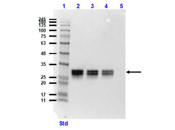
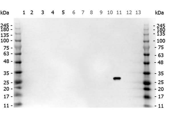
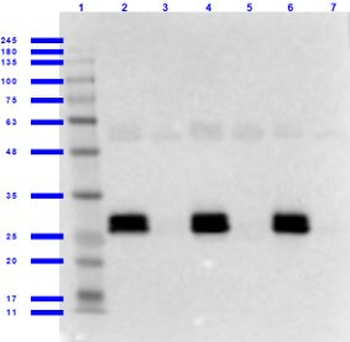
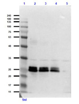
Western blot of Mouse Anti-GFP Antibody. Lane 1: Opal Prestained Molecular Weight Marker. Lane 2: HeLa WC Lysate+ GFP protein (p/n orb348668 [10 µg]/ p/n orb345957 [50 ng]). Lane 3: HeLa WC Lysate+ GFP protein (10 µg/20 ng). Lane 4: HeLa WC Lysate+ GFP protein (10 µg/10 ng). Lane 5: HeLa Whole Cell Lysate (p/n orb348668) (10 µg). Primary Antibody: Anti-GFP at 1:1000 overnight at 2-8°C. Secondary Antibody: Rabbit Anti-Mouse IgG HRP (p/n orb347544) at 1:40000 for 30 mins at RT. Block: BlockOut Buffer (p/n orb348644). Expected MW: ~27 kDa.
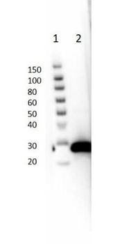
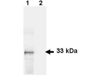
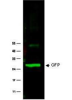
Western blot of Mouse Anti-GFP Antibody. Lane 1: Thermo SuperSignal Molecular Weight Marker. Lane 2: GFP protein (p/n orb345957) [50 ng]. Primary Antibody: Anti-GFP at 1:1000 overnight at 2-8°C. Secondary Antibody: Rabbit Anti-Mouse IgG HRP (p/n orb347544) at 1:40000 for 30 mins at RT. Block: BlockOut Buffer (p/n orb348644). Expected MW: ~27 kDa.
- Item 1 of 28
- Item 1 of 28
- Item 1 of 28
- Item 1 of 6
- Item 1 of 6


Submit a review
Filter by Rating

- 5 stars

- 4 stars
- 3 stars
- 2 stars
- 1 stars
