You have no items in your shopping cart.
Cart summary
Item 1 of 5
Item 1 of 5
GFAP Antibody
Catalog Number: orb1939391

| Catalog Number | orb1939391 |
|---|---|
| Category | Antibodies |
| Description | Mouse Monoclonal Antibody (Mab) |
| Species/Host | Mouse |
| Clonality | Monoclonal |
| Clone Number | 183CT3.1.5 |
| Tested applications | IF, IHC-P, WB |
| Reactivity | Human |
| Isotype | IgG2b,k |
| Dilution range | IF: 1:10~50, WB: 1:4000, WB: 1:500~1000, IHC-P-Leica: 1:1000, IHC: 1:2000 |
| Form/Appearance | Purified monoclonal antibody supplied in PBS with 0.09% (W/V) sodium azide. This antibody is purified through a protein G column, followed by dialysis against PBS. |
| Conjugation | Unconjugated |
| MW | 49880 Da |
| Target | This GFAP monoclonal antibody is generated from mouse immunized with GFAP recombinant protein. |
| UniProt ID | P14136 |
| NCBI | NP_001229305.1, NP_001124491.1, NP_002046.1 |
| Storage | Maintain refrigerated at 2-8°C for up to 2 weeks. For long term storage store at -20°C in small aliquots to prevent freeze-thaw cycles |
| Alternative names | Glial fibrillary acidic protein, GFAP, GFAP Read more... |
| Note | For research use only |
| Expiration Date | 12 months from date of receipt. |

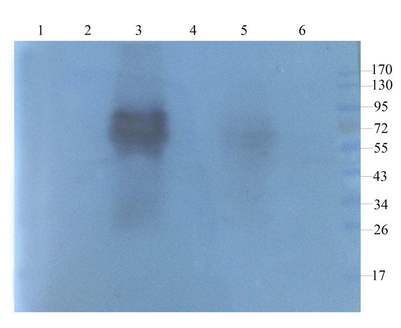
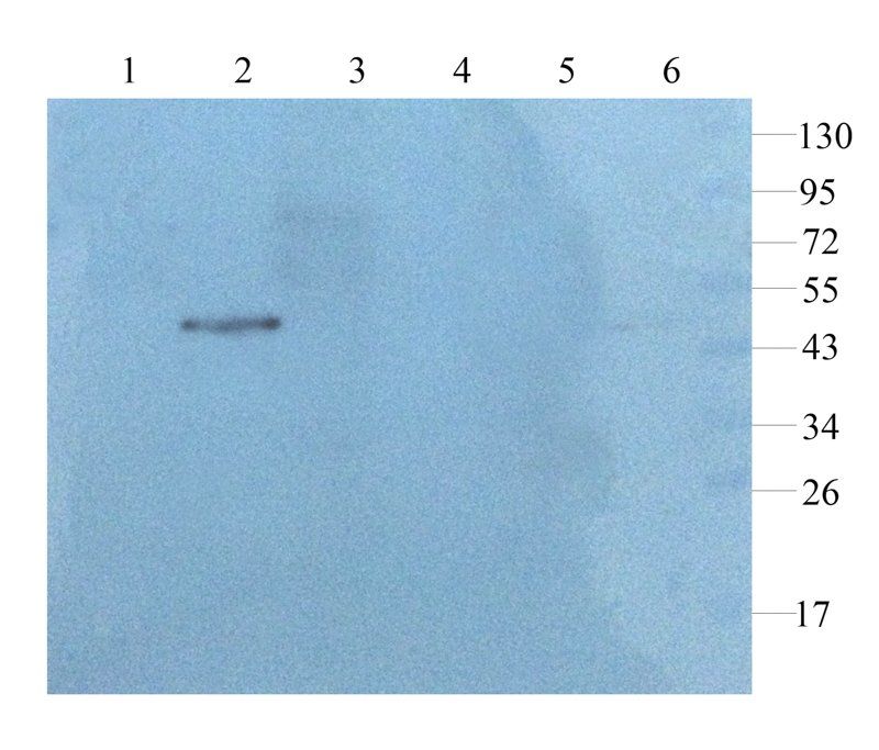
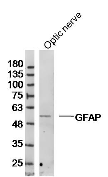
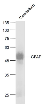
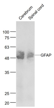
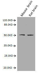
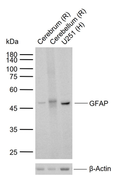
All lanes: Anti-GFAP Antibody at 1:4000 dilution. Lane 1: human brain lysate. Lane 2: human cerebellum lysate.Lysates/proteins at 20 µg per lane. Secondary Goat Anti-mouse IgG, (H+L), Peroxidase conjugated at 1/10000 dilution. Predicted band size: 50 kDa. Blocking/Dilution buffer: 5% NFDM/TBST.
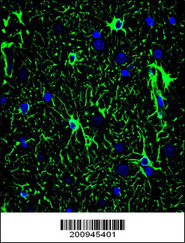
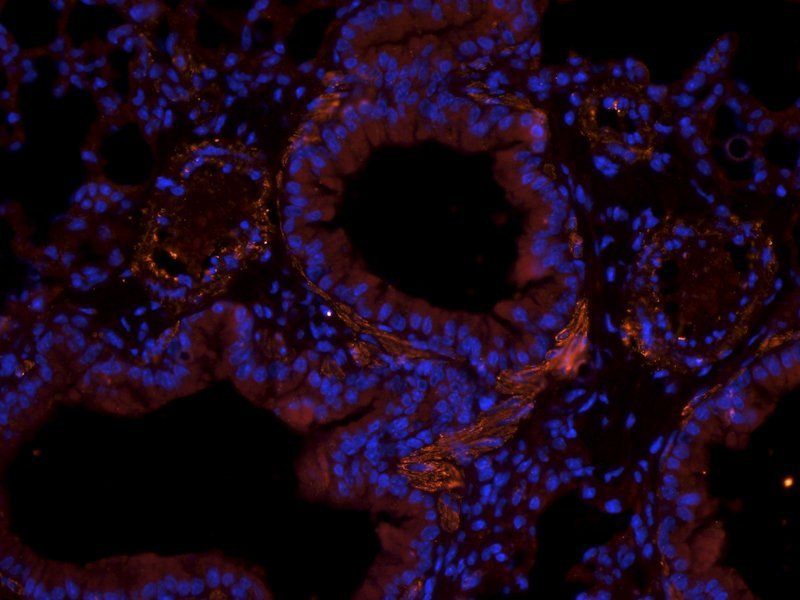
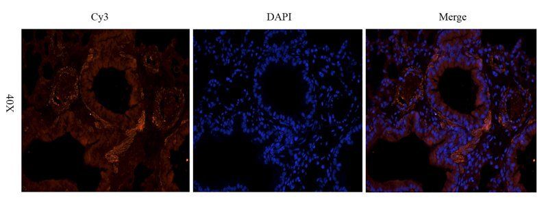
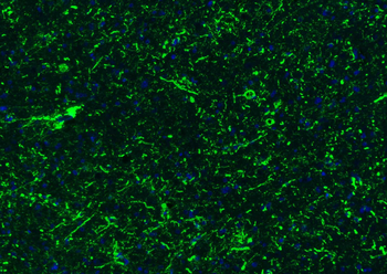
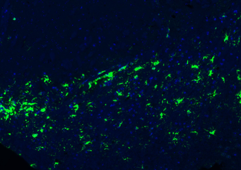
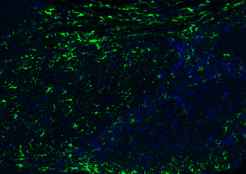
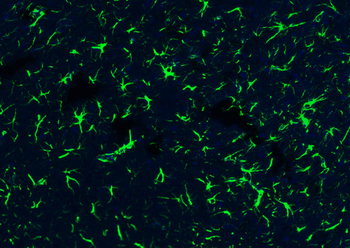
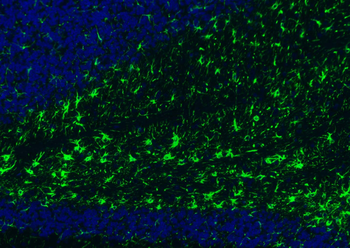
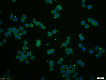
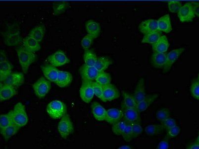
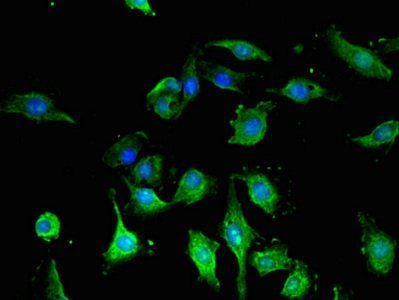
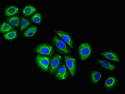
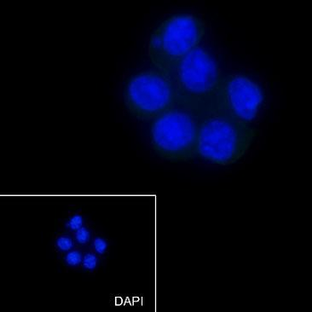
Confocal immunofluorescent analysis of GFAP Antibody with brain tissue followed by Alexa Fluor 488-conjugated goat anti-mouse lgG (green). DAPI was used to stain the cell nuclear (blue).
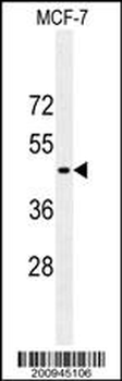
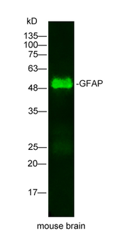
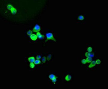
GFAP Antibody western blot analysis in MCF-7 cell line lysates (35 μg/lane). This demonstrates the GFAP antibody detected the GFAP protein (arrow).
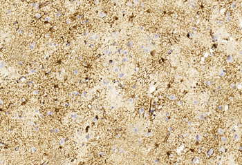
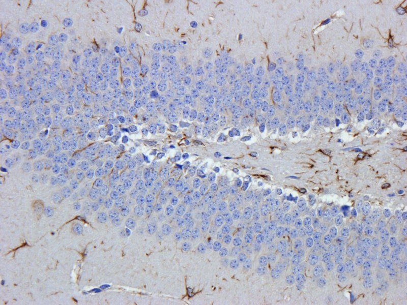
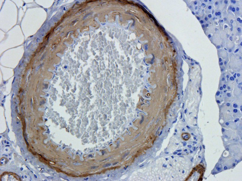
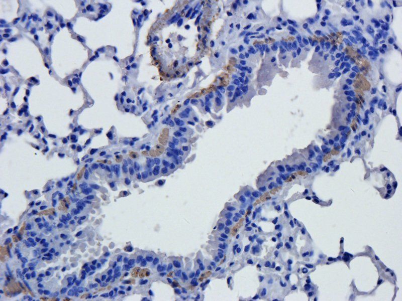
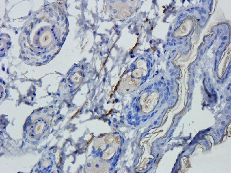
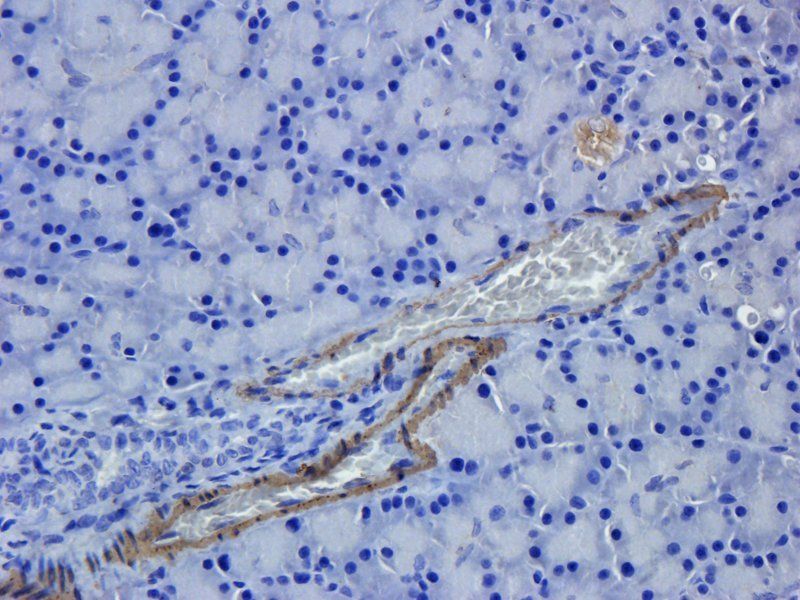
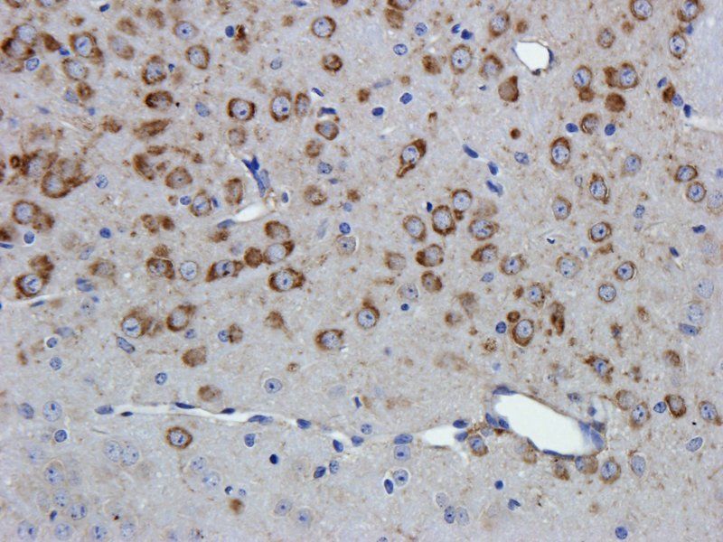
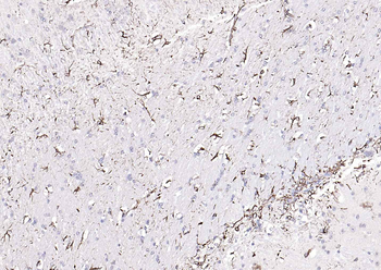
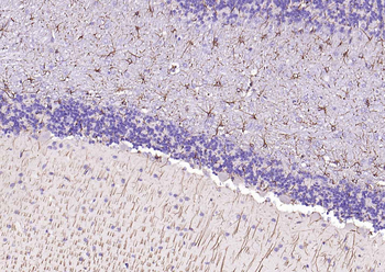
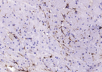
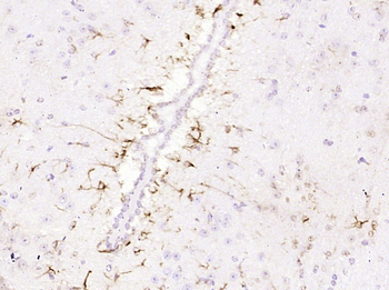
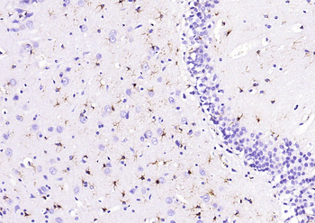
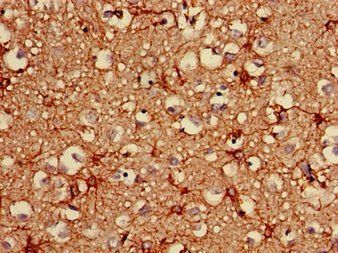
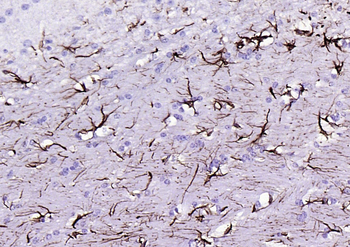
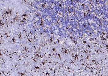
Immunohistochemical analysis of paraffin-embedded Human brain section using Pink1. diluted at 1:2000 dilution. A undiluted biotinylated goat polyvalent antibody was used as the secondary, followed by DAB staining.
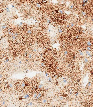
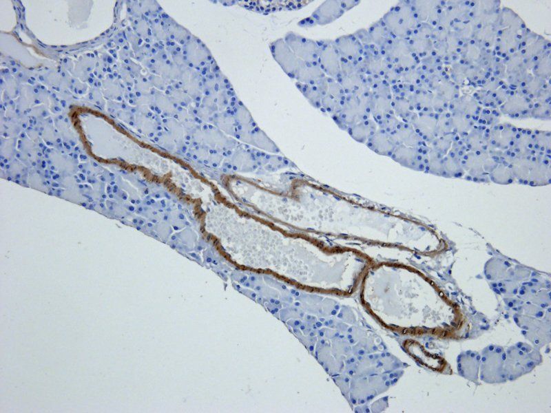
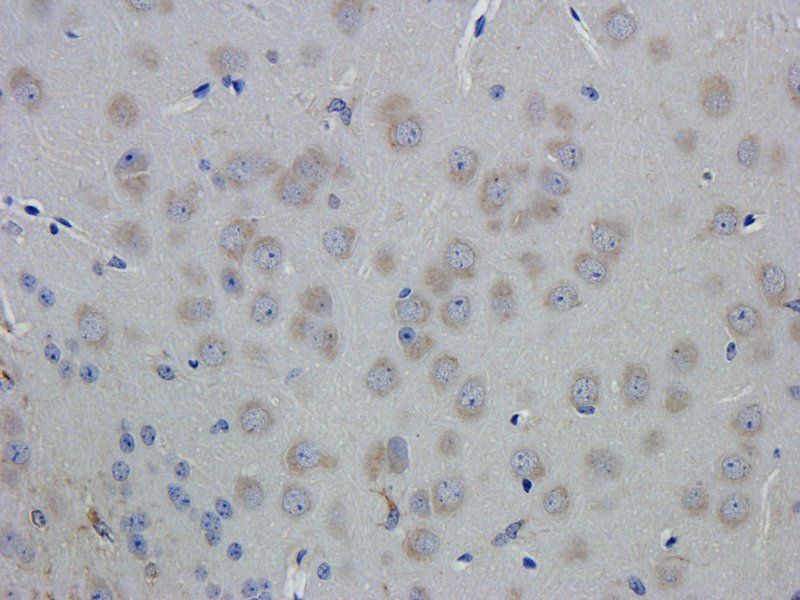
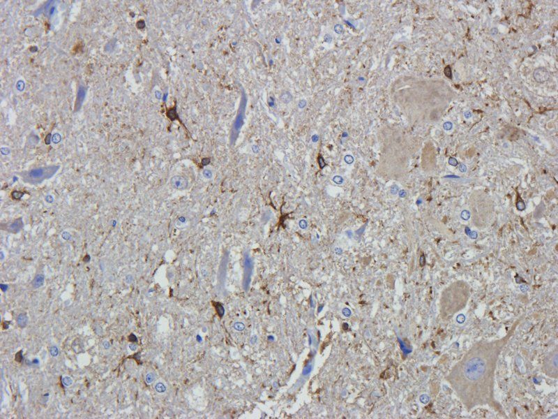
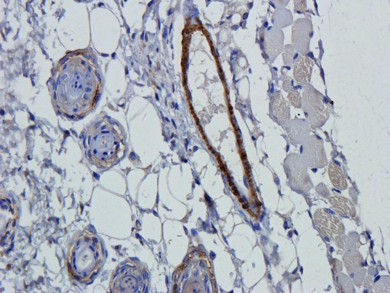
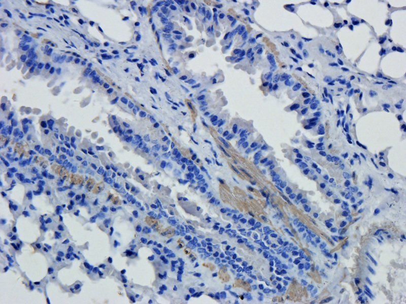
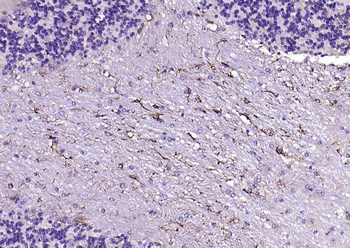
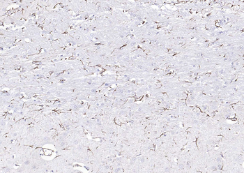
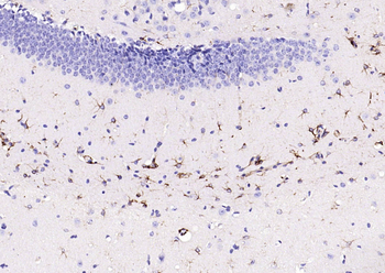
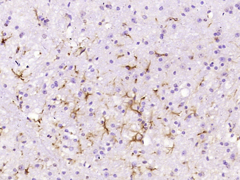
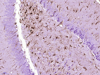
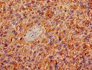
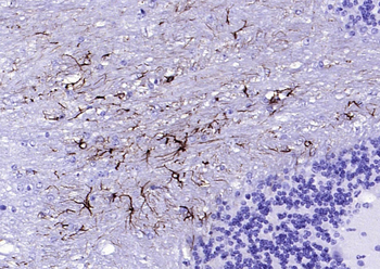
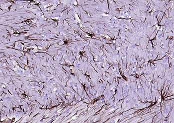
Immunohistochemical analysis of paraffin-embedded human brain tissue performed on the Leica BOND RXm. Tissue was fixed with formaldehyde at room temperature; antigen retrieval was by heat mediation with a EDTA buffer (pH9.0). Samples were incubated with primary antibody (1:1000) for 1 hours at room temperature. A undiluted biotinylated CRF Anti-Polyvalent HRP Polymer antibody was used as the secondary Antibody.
- Item 1 of 15
GFAP antibody [orb10706]
ELISA, ICC, IF, IHC-P, WB
Human, Mouse, Rat
Rabbit
Polyclonal
Unconjugated
100 μg - Item 1 of 12
GFAP Rabbit Polyclonal Antibody [orb500829]
FC, IF, IHC-Fr, IHC-P
Bovine, Canine, Porcine, Rabbit, Sheep
Human, Mouse, Rat
Rabbit
Polyclonal
Unconjugated
50 μl, 100 μl, 200 μl - Item 1 of 13
GFAP Mouse Monoclonal Antibody [orb500621]
ICC, IF, IHC-Fr, IHC-P, WB
Human, Rat
Human, Mouse, Rat
Mouse
Monoclonal
Unconjugated
200 μl, 100 μl, 50 μl - Item 1 of 9
GFAP Monoclonal Antibody [orb401780]
ELISA, FC, IF, IHC, WB
Human, Mouse, Rat
Mouse
Monoclonal
Unconjugated
100 μl, 50 μl - Item 1 of 7
GFAP Recombinant Rabbit Monoclonal Antibody [orb1152219]
IF, IHC-Fr, IHC-P, WB
Mouse, Rat
Human, Mouse, Rat
Rabbit
Recombinant
Unconjugated
100 μl, 50 μl