You have no items in your shopping cart.
Cart summary
Item 1 of 8
Item 1 of 8
GFAP Antibody
Catalog Number: orb1294403

| Catalog Number | orb1294403 |
|---|---|
| Category | Antibodies |
| Description | Rabbit polyclonal antibody to GFAP |
| Species/Host | Rabbit |
| Clonality | Polyclonal |
| Tested applications | IF, IHC-Fr, IHC-P, WB |
| Reactivity | Human, Mouse, Rabbit |
| Isotype | Rabbit-IgG |
| Immunogen | Synthetic peptide / encompassing a sequence within the C-terminus region. |
| Dilution range | Western Blot 1:1000-1:2000 Immunofluorescence 1:200 – 1:500 Immunohistochemistry (Frozen) 1:100 – 1:300 Immunohistochemistry (Paraffin) 1:100 – 1:300 |
| Conjugation | Unconjugated |
| Target | GFAP |
| Storage | Store at +4°C for short term storage. Long time storage is recommended at -20°C 100mM Tris Glycine, 1% BSA, 20% Glycerol (pH7). 0.025% ProClin 300 was added as a preservative |
| Note | For research use only |
| Expiration Date | 12 months from date of receipt. |
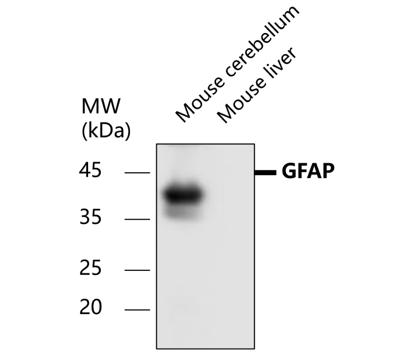
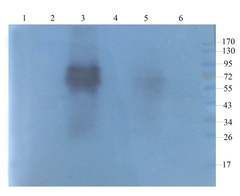
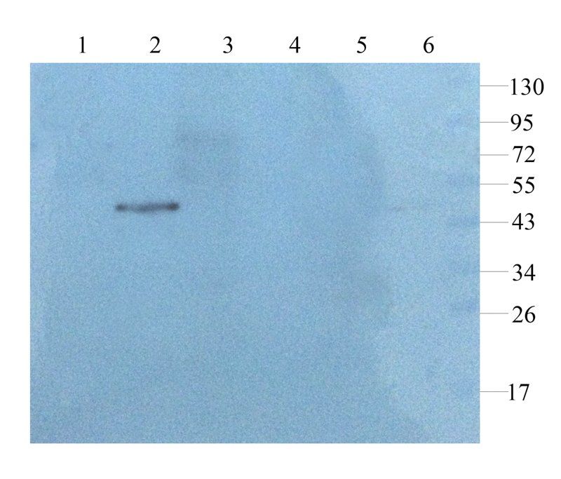
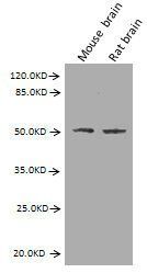
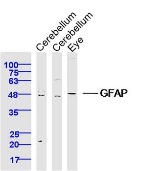
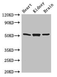
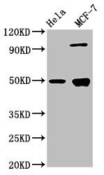
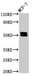
All lanes: Anti-GFAP antibody at 1/1000 dilution, Lysates/proteins at 60 μg per lane. This blot was produced using a 12% SDS-PAGE. Nitrocellulose was then blocked for an hour before being incubated with orb1294403 overnight at 4°C.
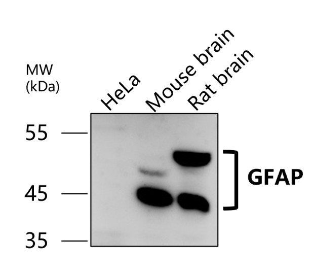
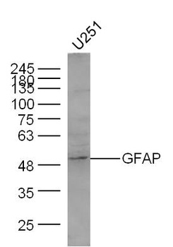
All lanes: Anti-GFAP antibody at 1/1000 dilution, Lysates/proteins at 60 μg per lane. This blot was produced using a 12% SDS-PAGE. Nitrocellulose was then blocked for an hour before being incubated with orb1294403 overnight at 4°C.
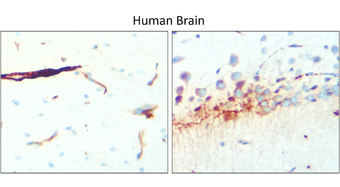
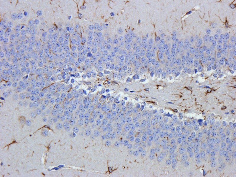
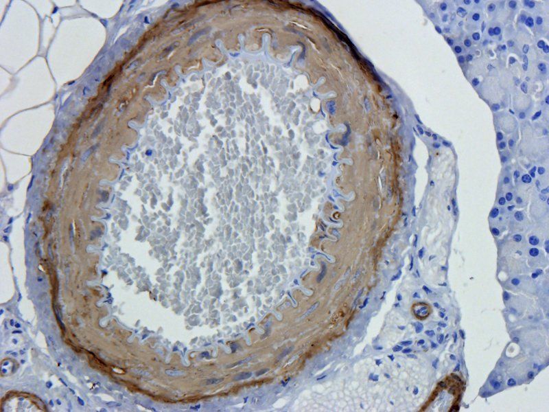
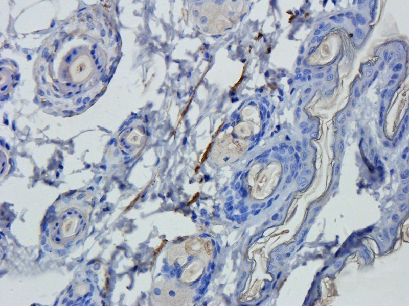
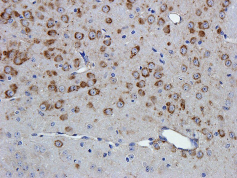
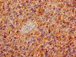
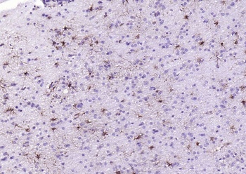
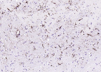
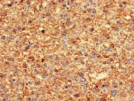
Immunohistochemical analysis of paraffin embedded Human brain tissue labeling GFAP antibody with orb1294403 at 1/100.
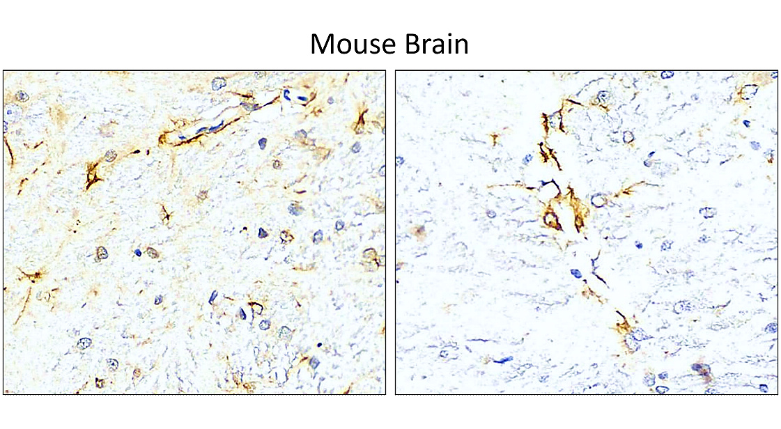
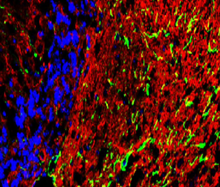
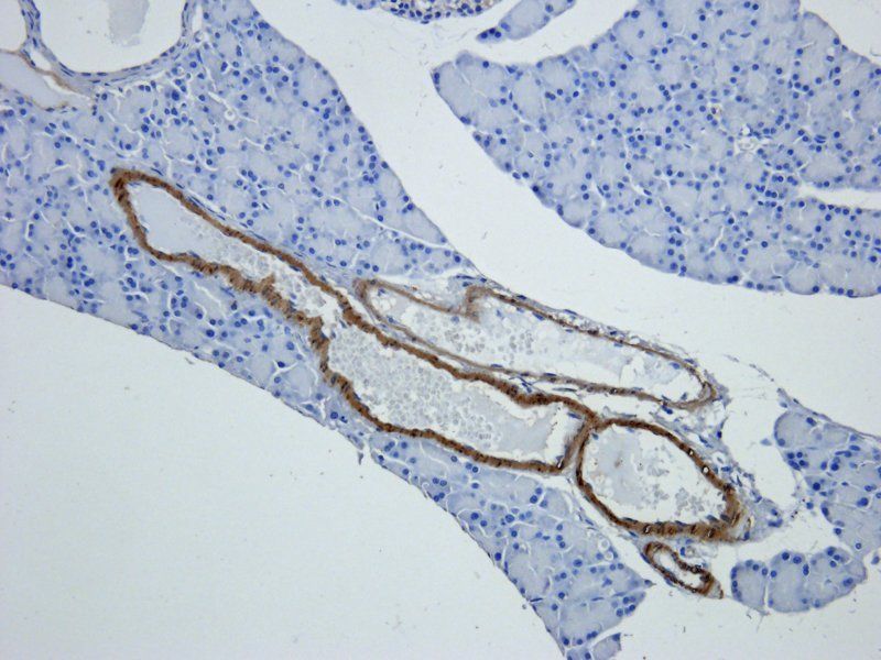
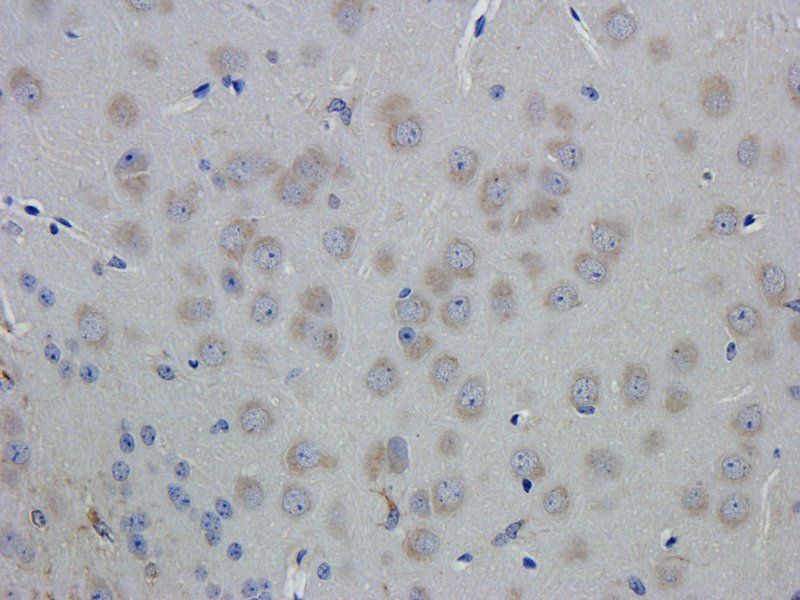
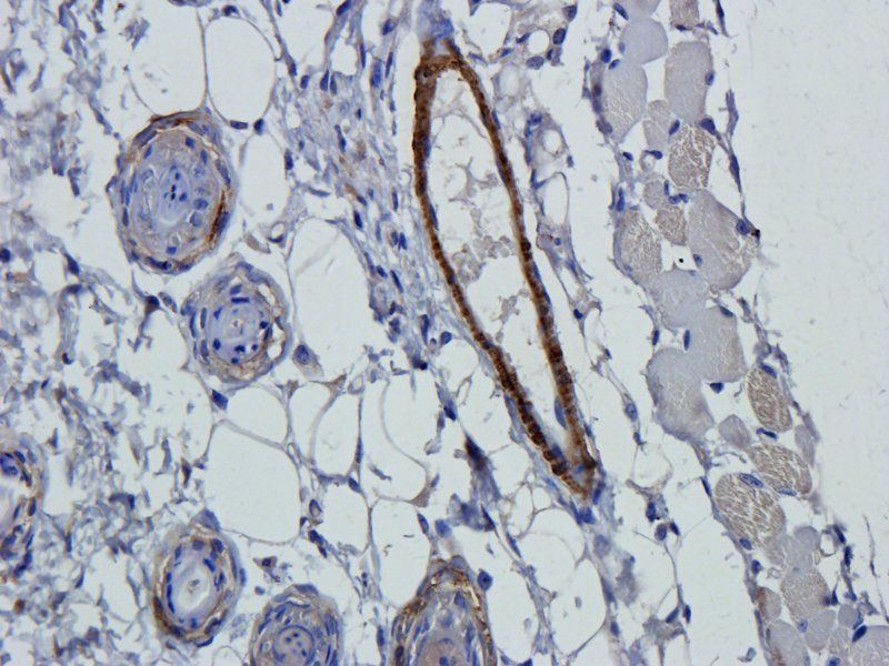
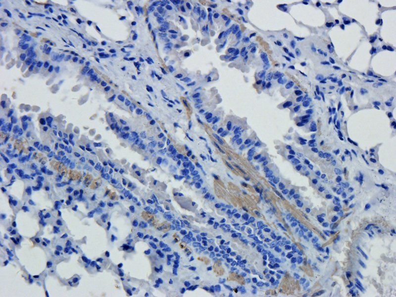
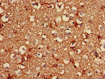
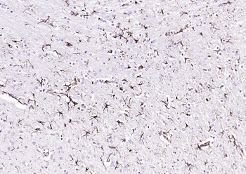
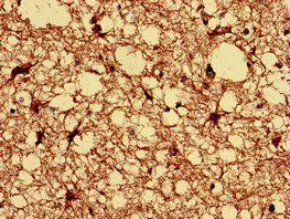
Immunohistochemical analysis of paraffin embedded Mouse brain tissue labeling GFAP antibody with orb1294403 at 1/100.
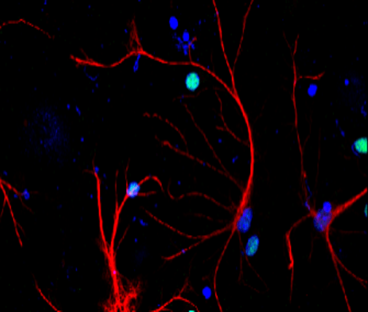
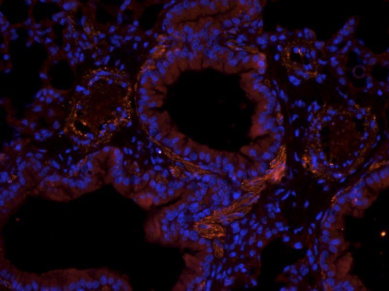
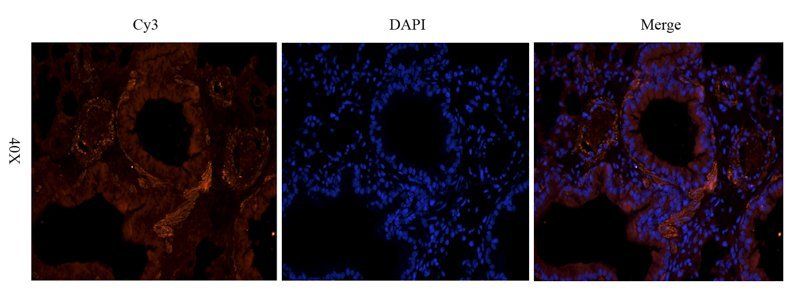
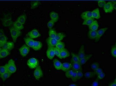
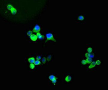
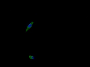
Immunofluorescent analysis. Sample: primary cortical neurons; Red: GFAP (orb1294403): 1-200; Blue: DAPI was used as the nuclear counter stain. Fixed: 4% paraformaldehyde at RT for 20 min.
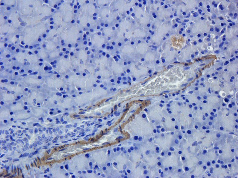
Immunohistochemical of frozen sections. Sample: mouse cerebellum. Green: GFAP (orb1294403): 1-200; Anti-rabbit 488: 1-500.
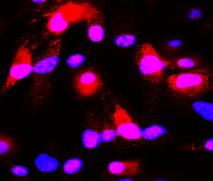
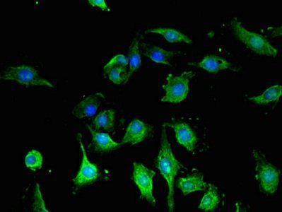
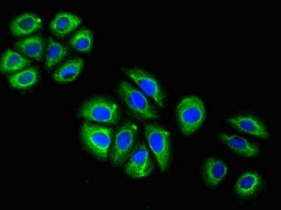
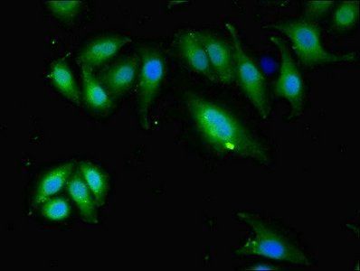
Immunofluorescence: cells were fixed with 4% paraformaldehyde for 10 min at RT, permeabilized with 0.1% NP-40 for 10 min at RT then blocked with 5% BSA for 30 min at room temperature. Cells were stained with orb1294403 anti-GFAP antibody (red) at 1:200 and 4°C. DAPI (blue) was used as the nuclear counter stain.
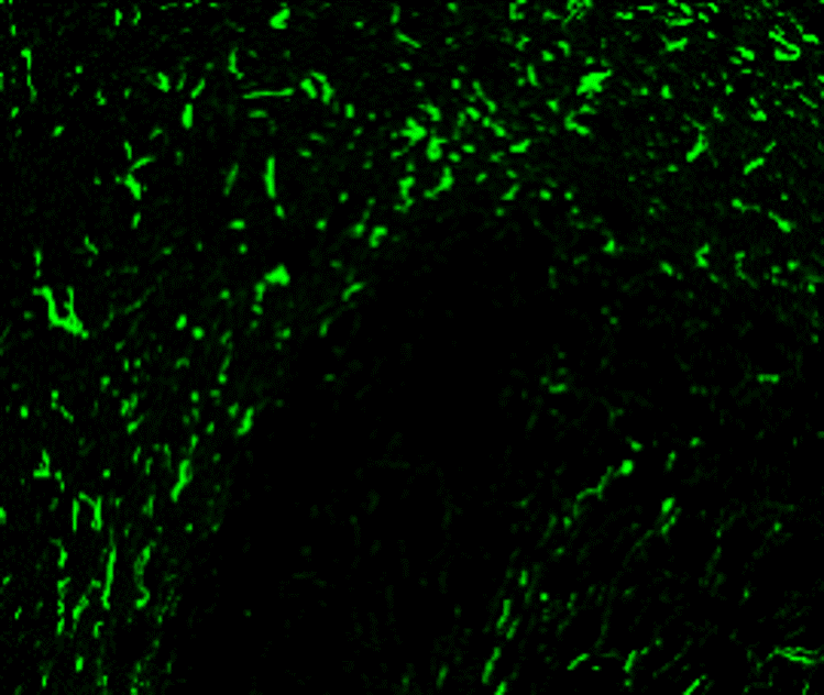
Immunohistochemical of frozen sections. Sample: mouse cerebellum. Green: GFAP (orb1294403): 1-200; Anti-rabbit 488: 1-500.
- Item 1 of 15
GFAP antibody [orb10706]
ELISA, ICC, IF, IHC-P, WB
Human, Mouse, Rat
Rabbit
Polyclonal
Unconjugated
100 μg, 200 μg - Item 1 of 9
GFAP antibody [orb401780]
ELISA, FC, IF, IHC, WB
Human, Mouse, Rat
Mouse
Monoclonal
Unconjugated
100 μl, 50 μl - Item 1 of 8
GFAP antibody [orb500829]
FC, IF, IHC-P, WB
Bovine, Canine, Porcine, Rabbit, Sheep
Human, Mouse, Rat
Rabbit
Polyclonal
Unconjugated
50 μl, 100 μl, 200 μl - Item 1 of 7
- Item 1 of 6
Glial fibrillary acidic protein antibody [orb240188]
ELISA, IF, IHC, WB
Human
Rabbit
Polyclonal
Unconjugated
50 μg, 100 μg


Submit a review
Filter by Rating

- 5 stars

- 4 stars
- 3 stars
- 2 stars
- 1 stars
