You have no items in your shopping cart.
Cart summary
Item 1 of 4
Item 1 of 4
FNIP1 Antibody: HRP
Catalog Number: orb55607

| Catalog Number | orb55607 |
|---|---|
| Category | Antibodies |
| Description | Rabbit polyclonal antibody against FNIP1 conjugated to HRP. |
| Species/Host | Rabbit |
| Clonality | Polyclonal |
| Tested applications | ELISA, IHC, WB |
| Reactivity | Human, Mouse, Rat |
| Immunogen | Synthetic peptide from the mid-protein of Human FNIP1 |
| Concentration | 1 mg/ml |
| Dilution range | WB (1:1000); ICC/IF (1:100); IHC (1:50) |
| Conjugation | HRP |
| MW | 130 kDa |
| Target | FNIP1 |
| Entrez | 96459 |
| UniProt ID | Q8TF40 |
| NCBI | NP_001008738.2 |
| Storage | Conjugated antibodies should be stored according to the product label |
| Buffer/Preservatives | 73.64mM Carbonate, 54.55mM Ethanolamine, 45.45mM Cyanoborohydride, 18.18mM Sodium Hydroxide and 0.23mM Citrate in dH2O |
| Alternative names | FNIP1 antibody, Folliculin-interacting protein 1 a Read more... |
| Note | For research use only |
| Application notes | A 1:1000 dilution of SPC-718 was sufficient for detection of FNIP1 in 15 µg of mouse kidney cell lysates by ECL immunoblot analysis using goat anti-rabbit IgG:HRP as the secondary antibody. |
| Expiration Date | 12 months from date of receipt. |

Immunocytochemistry/Immunofluorescence analysis using Rabbit Anti-FNIP1 Polyclonal Antibody. Tissue: C2C12 Cells (Mouse Myoblast cell line). Species: Mouse. Fixation: 4% Formaldehyde for 15 min at RT. Primary Antibody: Rabbit Anti-FNIP1 Polyclonal Antibody at 1:100 for 60 min at RT. Secondary Antibody: Goat Anti-Rabbit ATTO 488 at 1:200 for 60 min at RT. Counterstain: Phalloidin Texas Red F-Actin stain; DAPI (blue) nuclear stain at 1:1000, 1:5000 for 60 min at RT, 5 min at RT. Localization: Cytoplasm. Magnification: 60X. (A) DAPI (blue) nuclear stain (B) Phalloidin Texas Red F-Actin stain (C) FNIP1 Antibody (D) Composite.
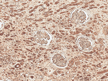
Immunohistochemistry analysis using Rabbit Anti-FNP1 Polyclonal Antibody. Tissue: Renal Cell Carcinoma. Species: Human. Fixation: Formalin Fixed Paraffin-Embedded. Primary Antibody: Rabbit Anti-FNP1 Polyclonal Antibody at 1:50 for 30 min at RT. Counterstain: Hematoxylin. Magnification: 10X. HRP-DAB Detection.
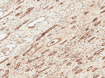
Immunohistochemistry analysis using Rabbit Anti-FNP1 Polyclonal Antibody. Tissue: Renal Cell Carcinoma. Species: Human. Fixation: Formalin Fixed Paraffin-Embedded. Primary Antibody: Rabbit Anti-FNP1 Polyclonal Antibody at 1:50 for 30 min at RT. Counterstain: Hematoxylin. Magnification: 10X. HRP-DAB Detection.
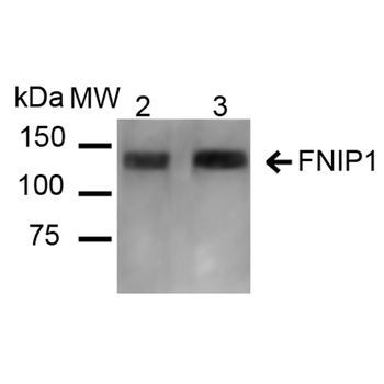
Western blot analysis of Mouse, Rat Kidney showing detection of ~131 kDa FNIP1 protein using Rabbit Anti-FNIP1 Polyclonal Antibody. Lane 1: Molecular Weight Ladder (MW). Lane 2: Mouse Kidney cell lysates. Lane 3: Rat Kidney cell lysates. Load: 20 μg. Block: 5% Skim Milk in 1X TBST. Primary Antibody: Rabbit Anti-FNIP1 Polyclonal Antibody at 1:1000 for 16 hours at 4°C. Secondary Antibody: Goat Anti-Rabbit IgG: HRP at 1:2000 for 60 min at RT. Color Development: ECL solution for 6 min at RT. Predicted/Observed Size: ~131 kDa.

FNIP1 Rabbit Polyclonal Antibody (HRP) [orb470827]
IHC-Fr, IHC-P
Bovine, Canine, Equine, Gallus, Human, Mouse, Sheep
Rat
Rabbit
Polyclonal
HRP
100 μlFNIP2 Rabbit Polyclonal Antibody (HRP) [orb470828]
WB
Bovine, Canine, Equine, Gallus, Mouse, Rat, Sheep
Human
Rabbit
Polyclonal
HRP
100 μl
