You have no items in your shopping cart.
Cart summary
Item 1 of 3
Item 1 of 3
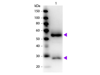
Fab Mouse IgG (H&L) Antibody
Catalog Number: orb348477

| Catalog Number | orb348477 |
|---|---|
| Category | Antibodies |
| Description | Fab Mouse IgG (H&L) antibody |
| Species/Host | Rabbit |
| Clonality | Polyclonal |
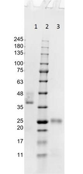
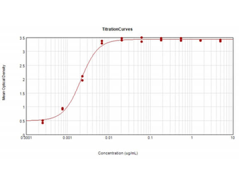
| Tested applications | ELISA, IHC, WB |
| Reactivity | Mouse |
| Isotype | IgG Fab |
| Immunogen | Mouse IgG whole molecule |
| Concentration | 1.0 mg/mL |
| Dilution range | ELISA: 1:20,000 - 1:100,000, IHC: 1:1,000 - 1:5,000, WB: 1:2,000 - 1:10,000 |
| Form/Appearance | Liquid (sterile filtered) |
| Purity | This product was prepared from monospecific antiserum by immunoaffinity chromatography using Mouse IgG coupled to agarose beads followed by solid phase adsorption(s) to remove any unwanted reactivities, papain digestion and chromatographic separation. Assay by immunoelectrophoresis resulted in a single precipitin arc against anti-Rabbit Serum. No reaction was observed against anti-Papain or anti-Rabbit IgG F(c). |
| Conjugation | Unconjugated |
| Storage | Store vial at 4° C prior to opening. This product is stable at 4° C as an undiluted liquid. Dilute only prior to immediate use. |
| Buffer/Preservatives | 0.01% (w/v) Sodium Azide |
| Alternative names | Rabbit Fab Anti-Mouse IgG Antibody, Rabbit Fab Fra Read more... |
| Note | For research use only |
| Application notes | Suitable for highly specific immunological methods requiring extremely low background levels, absence of F(c) mediated binding, lot-to-lot consistency, high titer and specificity. |
| Expiration Date | 12 months from date of receipt. |

CASPR1 is located pre-synaptically in close vicinity to the synaptic ribbon. A) High resolution confocal analysis of rod photoreceptor synapses in the OPL of the mouse retina (0.5 µm-thin sections) that were triple-immunolabelled with the rabbit polyclonal antibody against CASPR1, mouse monoclonal antibody 2D9 against RIBEYE and the indicated third primary antibodies (A-F). In (A), the outline of a single presynaptic terminal was visualized by immunolabelling with antibodies against PSD95 (A2). The CASPR1 immunosignal is located within the presynaptic terminal in close vicinity to the synaptic ribbon. Furthermore, presynaptic CASPR1 was found in close vicinity to the active zone markers RIM2 (B) and CASK (C, D, E). In contrast, the CASPR1 signal did not overlap with the postsynaptic signal for mGluR6 that is localized at the tip of invaginating ON-bipolar cells that are located also in close vicinity to the synaptic ribbon (Appendix Fig. S8F). Appendix Figs. S8B, C, D, F were obtained by confocal microscopy; Appendix Figs. S8A, E by super-resolution structured illumination-microscopy (SR-SIM). OPL, outer plexiform layer; INL, inner nuclear layer; IPL, inner plexiform layer. Scale bars: 1 µm (A-F).
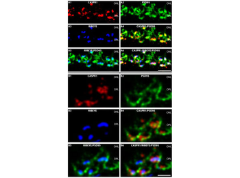
Confocal images of semi-thin sections of the mouse retina triple-immunolabelled with mouse monoclonal antibodies against CASPR1, rabbit polyclonal antibodies against PSD-95 (L667) and rabbit polyclonal antibodies against RIBEYE (U2656), as described in the Materials and Methods section. In A4, A4 and B4, B5 the indicated two immunosignals are overlayered on each other; in A6, B6, all three immunosignals were overlayered on each other. ONL, outer nuclear layer; OPL, outer plexiform layer. Scale bars: 2 µm.
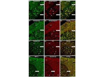
Semi-thin (0.5 µm-thin) sections of the mouse cerebellum double-immunolabeled with the monoclonal dynamin1xb antibody and the indicated other primary antibodies. The other primary antibodies against synaptotagmin1 (A, B), synaptic vesicle protein 2 (SV2; C) and RIM1/2 (D) were applied to label the synapses in order to better relate the dynamin1xb immunosignals to the synaptic regions. We observed a strong dynamin1xb immunosignal in the cerebellar cortex whereas the cerebellar medulla (white matter) that contains predominantly fiber tracts (but no synapses) was not immunolabeled. In the cerebellar cortex, dynamin1xb was highly enriched in the synaptic regions, i.e., the molecular layer (mol) of the cerebellar cortex and the giant synapses in the granule cell layer (arrows) of the cerebellar cortex. No significant dynamin1xb immunosignal was observed in the medulla of the cerebellum that predominantly contains axonal fiber tracts. (A, B, D) was obtained by epifluorescence microscopy; (C) was obtained by confocal microscopy. Abbreviations: mol, molecular layer; Pu, Purkinje cell layer; gr, granule cell layer. Scale bars: 50 µm (A–D).
- Item 1 of 2
- Item 1 of 1
Fab Mouse IgG (H&L) Antibody Rhodamine Conjugated [orb348470]
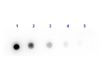
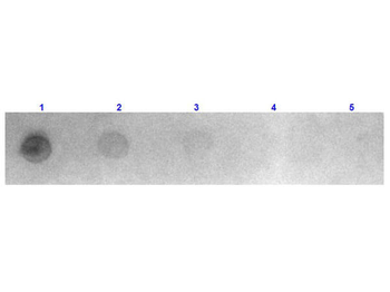
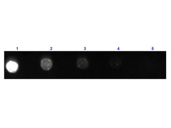
DOT, FC, FLISA, IF
Mouse
Goat
Polyclonal
TRITC
1 mg - Item 1 of 1
Fab Mouse IgG (H&L) Antibody Fluorescein Conjugated [orb348472]
DOT, IF, WB
Mouse
Goat
Polyclonal
FITC
1 mg - Item 1 of 1
Fab Mouse IgG (H&L) Antibody Peroxidase Conjugated [orb348473]
ELISA, IHC, WB
Mouse
Goat
Polyclonal
HRP
1 mg - Item 1 of 1
Fab Mouse IgG (H&L) Antibody Biotin Conjugated [orb348474]
DOT, ELISA, IHC, WB
Mouse
Goat
Polyclonal
Biotin
1 mg