You have no items in your shopping cart.
Cart summary
Item 1 of 1
ESET antibody
Catalog Number: orb345472

| Catalog Number | orb345472 |
|---|---|
| Category | Antibodies |
| Description | ESET antibody |
| Species/Host | Rabbit |
| Clonality | Polyclonal |
| Tested applications | ELISA, IHC, WB |
| Reactivity | Human |
| Isotype | IgG |
| Immunogen | This affinity purified antibody was prepared from whole rabbit serum produced by repeated immunizations with a synthetic peptide corresponding to amino acids 1050-1075 of human ESET. |
| Concentration | 1.17 mg/mL |
| Dilution range | ELISA: 1:10,000 - 1:50,000, IHC: User Optimized, WB: 1:1,000 - 1:2,500 |
| Form/Appearance | Liquid (sterile filtered) |
| Purity | This is an affinity purified antibody produced by immunoaffinity chromatography using the immunizing peptide after immobilization to a solid phase. Reactivity occurs against human ESET protein. |
| Conjugation | Unconjugated |
| UniProt ID | Q15047 |
| NCBI | 224177469 |
| Storage | Store vial at -20° C prior to opening. Aliquot contents and freeze at -20° C or below for extended storage. Avoid cycles of freezing and thawing. Centrifuge product if not completely clear after standing at room temperature. This product is stable for several weeks at 4° C as an undiluted liquid. Dilute only prior to immediate use. |
| Buffer/Preservatives | 0.01% (w/v) Sodium Azide |
| Alternative names | rabbit anti-ESET antibody, ERG-associated protein Read more... |
| Note | For research use only |
| Application notes | This affinity purified antibody has been tested for use in ELISA and western blotting. Western blotting shows reactivity specific for human ESET detecting a band at approximately 170 kDa. Specific band detection by western blot is blocked by peptide competition by pre-incubating the antibody with the immunizing peptide prior to reaction with the membrane. Reactivity in other immunoassays is unknown. |
| Expiration Date | 12 months from date of receipt. |
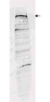
Western blot analysis is shown using Biorbyt's Affinity Purified anti-ESET antibody to detect human ESET present in a 293 whole cell lysate (p/n orb348669). ~30 µg of lysate was loaded per lane for SDS-PAGE. Comparison to a molecular weight marker (not shown) indicates a single band of ~170 kDa is detected. Peptide competition (not shown) blocks staining of this band. The blot was incubated with a 1:1000 dilution of the antibody at room temperature for 2 h followed by detection using IRDye™800 labeled Goat-a-Rabbit IgG [H&L] diluted 1:5000 for 45 min.
- Item 1 of 2
SETDB1 antibody [orb341002]
IH, WB
Human, Mouse, Rat
Rabbit
Polyclonal
Unconjugated
200 μl, 100 μl, 50 μl - Item 1 of 2
- Item 1 of 2
- Item 1 of 2
- Item 1 of 1


Submit a review
Filter by Rating

- 5 stars

- 4 stars
- 3 stars
- 2 stars
- 1 stars
