You have no items in your shopping cart.
Cart summary
Item 1 of 6
Item 1 of 6
Erk1/2 Antibody: Biotin
Catalog Number: orb151496

| Catalog Number | orb151496 |
|---|---|
| Category | Antibodies |
| Description | Rabbit polyclonal to Erk1 (Biotin). The extracellular signal-regulated kinases 1 and 2 (ERK1 and ERK2), also called p44 and p42 MAP kinases, are members of the Mitogen Activated Protein Kinase (MAPK) family of proteins found in all eukaryotes. Because the 44 kDa ERK1 and the 42 kDa ERK2 are highly homologous and both function in the same protein kinase cascade, the two proteins are often referred to collectively as ERK1/2 or p44/p42 MAP kinase. They are both located in the cytosol and mitochondria. While the role of cytosol ERK1/2 is well studied and involved in multiple cellular functions, the role of mitochondrial ERK1/2 remains poorly understood. Both ERK 1 and 2 are activated by MEK1 or MEK2, by dual phosphorylation of a threonine and tyrosine residue in the activation loop (TEY motif) (1, 3). Either phosphorylation alone can induce an electrophoretic mobility shift, but both are required for activation of the kinase. This dual phosphorylation is efficiently detected by phosphorylation state-specific antibody directed to the pTEpY motif. Once activated, MAP kinases phosphorylate a broad spectrum of substrates, including cytoskeletal proteins, translation regulators, transcription factors, and the Rsk family of protein kinases. ERK1/2 activation is generally thought to confer a survival advantage to cells; however there is increasing evidence that suggests that the activation of ERK1/2 also contributes to cell death under certain conditions. ERK1/2 also is activated in neuronal and renal epithelial cells upon exposure to oxidative stress and toxicants or deprivation of growth factors, and inhibition of the ERK pathway blocks apoptosis.. |
| Species/Host | Rabbit |
| Clonality | Polyclonal |
| Tested applications | ELISA, ICC, IF, IHC, WB |
| Reactivity | Bovine, Drosophila, Frog, Gallus, Human, Mouse, Rat, Sheep |
| Immunogen | A 35 residue synthetic peptide, corresponding to Erk1 MAP kinase with the CGG spacer group added and the peptide coupled to KLH. |
| Concentration | 1 mg/ml |
| Dilution range | WB (1:1000), IHC (1:100), ICC/IF (1:100), FCM (1:100) |
| Conjugation | Biotin |
| MW | 42kDa (ERK2). |
| Target | ERK1 |
| Entrez | 50689 |
| UniProt ID | P21708 |
| NCBI | NP_059043.1 |
| Storage | Conjugated antibodies should be stored according to the product label |
| Buffer/Preservatives | 136.36mM Ethanolamine, and 9.55mM Sodium Bicarbonate in 95.45% PBS |
| Alternative names | ERK1 antibody, ERK2 antibody, ERT1 antibody, ERT2 Read more... |
| Note | For research use only |
| Application notes | A 1:1000 dilution of SPC-120 was sufficient for detection of ERK1/2 in 20 µg of HeLa cell lysate by ECL immunoblot analysis. |
| Expiration Date | 12 months from date of receipt. |

Immunocytochemistry/Immunofluorescence analysis using Rabbit Anti-Erk1/2 Polyclonal Antibody. Tissue: Cervical cancer cell line (HeLa). Species: Human. Fixation: 2% Formaldehyde for 20 min at RT. Primary Antibody: Rabbit Anti-Erk1/2 Polyclonal Antibody at 1:100 for 12 hours at 4°C. Secondary Antibody: APC Goat Anti-Rabbit (red) at 1:200 for 2 hours at RT. Counterstain: DAPI (blue) nuclear stain at 1:40000 for 2 hours at RT. Localization: Cytoplasm. Nucleus. Magnification: 100x. (A) DAPI (blue) nuclear stain. (B) Anti-Erk1/2 Antibody. (C) Composite.
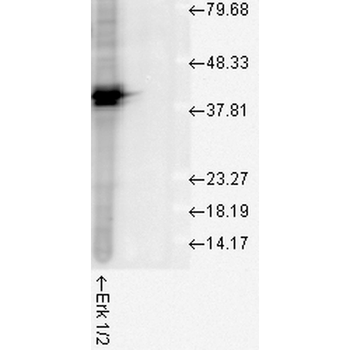
Western blot analysis of Human Cell line lysates showing detection of ERK1 protein using Rabbit Anti-ERK1 Polyclonal Antibody. Load: 15 μgprotein. Block: 1.5% BSA for 30 minutes at RT. Primary Antibody: Rabbit Anti-ERK1 Polyclonal Antibody at 1:1000 for 2 hours at RT. Secondary Antibody: Donkey Anti-Rabbit IgG: HRP for 1 hour at RT.
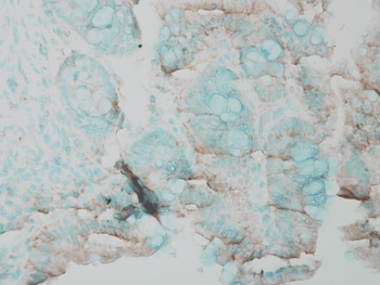
Immunohistochemistry analysis using Rabbit Anti-ERK1 Polyclonal Antibody. Tissue: Inflamed colon. Species: Mouse. Fixation: Formalin. Primary Antibody: Rabbit Anti-ERK1 Polyclonal Antibody at 1:25000 for 12 hours at 4°C. Secondary Antibody: Biotin Goat Anti-Rabbit at 1:2000 for 1 hour at RT. Counterstain: Methyl Green at 200uL for 2 min at RT.
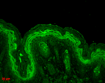
Immunohistochemistry analysis using Rabbit Anti-ERK1 Polyclonal Antibody. Tissue: backskin. Species: Mouse. Fixation: Bouin's Fixative Solution. Primary Antibody: Rabbit Anti-ERK1 Polyclonal Antibody at 1:100 for 1 hour at RT. Secondary Antibody: FITC Goat Anti-Rabbit (green) at 1:50 for 1 hour at RT. Localization: Cytoplasm.
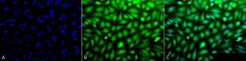
Immunocytochemistry/Immunofluorescence analysis using Rabbit Anti-Erk1/2 Polyclonal Antibody. Tissue: Cervical cancer cell line (HeLa). Species: Human. Fixation: 2% Formaldehyde for 20 min at RT. Primary Antibody: Rabbit Anti-Erk1/2 Polyclonal Antibody at 1:100 for 12 hours at 4°C. Secondary Antibody: FITC Goat Anti-Rabbit (green) at 1:200 for 2 hours at RT. Counterstain: DAPI (blue) nuclear stain at 1:40000 for 2 hours at RT. Localization: Cytoplasm. Nucleus. Magnification: 20x. (A) DAPI (blue) nuclear stain. (B) Anti-Erk1/2 Antibody. (C) Composite.
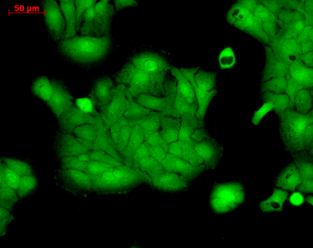
Immunocytochemistry/Immunofluorescence analysis using Rabbit Anti-ERK1 Polyclonal Antibody. Tissue: HaCaT cells. Species: Human. Fixation: Cold 100% methanol at -20°C for 10 minutes. Primary Antibody: Rabbit Anti-ERK1 Polyclonal Antibody at 1:100 for 12 hours at 4°C. Secondary Antibody: FITC Goat Anti-Rabbit at 1:50 for 1-2 hours at RT in dark. Localization: Cytoplasm. Nucleus.
