You have no items in your shopping cart.
Cart summary
Item 1 of 5
Item 1 of 5
Epitope Tag Antibody Sampler Kit
Catalog Number: orb342265

| Catalog Number | orb342265 |
|---|---|
| Category | Assays and Kits |
| Description | Epitope Tag Antibody Sampler Kit |
| Tested applications | ChIP, ELISA, FC, IF, IHC, IP, WB |
| Immunogen | GFP, GST, DYKDDDDK peptide (the same epitope recognized by Sigma's Anti-FLAG antibodies), 6X His peptide |
| Dilution range | ELISA: User Optimized, ChIP: User Optimized, FC: User Optimized, IHC: User Optimized, IF: User Optimized, IP: User Optimized, WB: User Optimized |
| Purity | 6X HIS antibody, GFP Antibody, DYKDDDDK Antibody (Binds to same epitope as Sigma's Anti-FLAG M2 Antibody), GST Antibody and Conjugated Secondary Antibodies are all affinity purified. |
| Conjugation | Unconjugated |
| Storage | Store Epitope Tag Kit at -20° C prior to opening. Aliquot antibodies and freeze at -20° C or below for extended storage. Avoid cycles of freezing and thawing. Centrifuge product if not completely clear after standing at room temperature. This product is stable for several weeks at 4° C as an undiluted liquid. Dilute only prior to immediate use. |
| Alternative names | FLAG, Green Fluorescent Protein, GFP, GST, six his Read more... |
| Note | For research use only |
| Application notes | Epitope Tag kit contains: 6X HIS Antibody 100 µg DYKDDDDK Antibody (Binds to same epitope as Sigma's Anti-FLAG M2 Antibody) 250 µg GFP Antibody 100 µg GST Antibody 1.0mg Anti-RABBIT IgG (H&L) (GOAT) Antibody Peroxidase Conjugated 100 µg Anti-GOAT IgG (H&L) (RABBIT) Antibody Peroxidase Conjugated 100 µg |
| Expiration Date | Please enquire. |
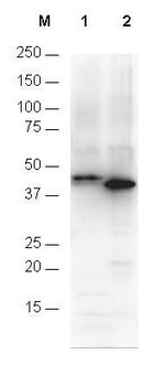
Anti-6X His epitope tag polyclonal antibody (orb345394) detects His-tagged recombinant proteins by western blot. The blot was blocked with 3% BSA in TBST for 45 min at RT. Antibody was incubated with blot at a 1:1000 dilution in TBST with 3% BSA for 1 hour at RT. Detection occurred using HRP Gt-a-Rabbit IgG diluted 1:80, 000 in blocking buffer (p/n orb348637) for 30 min at RT. Lane 1 was loaded with 12-Epitope Tag Protein Marker Lysate (p/n orb348609) which has the His epitope tag incorporated through a C-terminal linkage (~42 kDa). Lane 2 was loaded with His-SUMO-GFP recombinant protein which has the His epitope tag incorporated through an N-terminal linkage (~40 kDa). A 4-20% gradient gel was used to resolve the protein by SDS-PAGE. Proteins were transferred to nitrocellulose using standard methods. Molecular weights were estimated by comparison to standards (lane M).
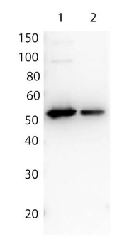
Biorbyt Affinity Purified anti FLAG™ Polyclonal Antibody (orb345396) detects both C terminal linked and N terminal linked FLAG™ tagged recombinant proteins by western blot. This antibody was used at a dilution of 1:2500 to detect 1.0 ?g of recombinant protein containing either the FLAG™ epitope tag linked at the carboxy (C) or the amino (N) terminus of the recombinant protein. A 4-20% gradient gel was used to resolve the protein by SDS-PAGE. The protein was transferred to nitrocellulose using standard methods. After blocking, the membrane was probed with the primary antibody for 1 h at room temperature followed by washes and reaction with a 1:10000 dilution of IRDye® 800 conjugated Gt-a-Rabbit IgG (H&L) MX10 for 30 min at room temperature.
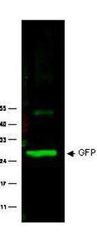
Biorbyt anti GFP polyclonal antibody (orb345367) was used to detect GFP protein. Wild type GFP (0.1 µg) was used to spike 30 µg of a HeLa whole cell lysate. This antibody detects a 27 kDa band corresponding to the epitope tag GFP. A 4-20% Tris-Glycine gradient gel was used for SDS-PAGE. The protein was transferred to nitrocellulose using standard methods. After blocking with 5% BLOTTO in PBS, the membrane was probed overnight at 4°C with the primary antibody diluted in 5% BLOTTO to 1:1000, followed by washes and reaction with a 1:10000 dilution of IRDye® 800 conjugated Goat-a-Rabbit IgG [H&L] MX10.
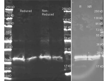
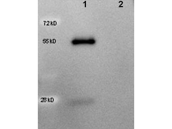
Biorbyt's anti-GST polyclonal antibody (orb345284) in western blot shows detection of recombinant GST (indicated by band at ~ 28 kDa). The SDS-PAGE contained approximately 0.2 µg of rGST loaded on to a 4-20% gradient gel for separation. After electrophoresis, the gel was transferred to nitrocellulose and blocked with "Blocking Buffer for Fluorescent Western Blotting" p/n orb348637 in TBS for 1 h at RT. The membrane was probed with anti-GST antibody at a 1:2000 dilution in blocking reagent, overnight at 4°C. For detection DyLight™800 conjugated Donkey-a-Goat IgG was used at a 1:20000 dilution (in blocking reagent) for 30 min at 25°C. Fluorescent data was collected on a LICOR Odyssey instrument.
Western Blot of HRP anti-Goat IgG antibody (orb346993) showing detection of 50 ng of goat IgG (lane 1) but not human IgG (lane 2). Samples were separated by 4-20% SDS-PAGE under reducing conditions and transferred to nitrocellulose membrane. The blot was blocked overnight at 4°C in 5% BSA in TBS. A 1:5000 dilution of antibody in Blocking Buffer for Fluorescent Western Blotting (p/n orb348637) was used to probe the membrane at room temperature for 1 h. The image was developed using Chemiluminescent FemtoMax™ Super Sensitive HRP Substrate for one minute.
Submit a review
Filter by Rating

- 5 stars

- 4 stars
- 3 stars
- 2 stars
- 1 stars
