You have no items in your shopping cart.
Cart summary
Item 1 of 7
Item 1 of 7
EpCAM Antibody
Catalog Number: orb749368

| Catalog Number | orb749368 |
|---|---|
| Category | Antibodies |
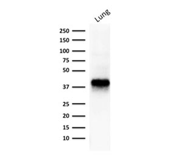
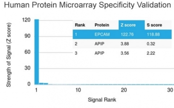
| Description | Epithelial cellular adhesion molecule (EpCAM), is also identified as epithelial specific antigen (ESA) and the 40kDa transmembrane epithelial glycoprotein EGP40. EpCAM is expressed on the baso-lateral cell surface in most simple epithelia and a vast majority of carcinomas. The VU-1D9 antibody has been used to distinguish adenocarcinoma from pleural mesothelioma and hepatocellular carcinoma. EpCAM antibody is also useful in distinguishing serous carcinomas of the ovary from mesothelioma. |
| Species/Host | Mouse |
| Clonality | Monoclonal |
| Clone Number | VU-1D9 |
| Tested applications | FACS, IF, IHC-P |
| Reactivity | Human |
| Isotype | Mouse IgG1, kappa |
| Immunogen | Small cell lung carcinoma cells were used as the immunogen for this EpCAM antibody VU-1D9. |
| Dilution range | Flow cytometry: 0.5-1ug/million cells,Immunofluorescence: 1-2ug/ml,Immunohistochemistry (FFPE): 0.5-1ug/ml for 30 min at RT |
| Purity | Protein G affinity chromatography |
| Conjugation | Unconjugated |
| Formula | 0.2 mg/ml in 1X PBS with 0.1 mg/ml BSA (US sourced) and 0.05% sodium azide |
| Hazard Information | This EpCAM antibody is available for research use only. |
| Entrez | 4072 |
| Storage | Store the EpCAM antibody at 2-8°C (with azide) or aliquot and store at -20°C or colder (without azide). |
| Buffer/Preservatives | 0.2 mg/ml in 1X PBS with 0.1 mg/ml rAlbumin (US sourced) and 0.05% sodium azide |
| Note | For research use only |
| Application notes | The concentration stated for each application is a general starting point. Variations in protocols, secondaries and substrates may require the EpCAM antibody to be titered up or down for optimal performance.1. Staining of formalin/paraffin tissues REQUIRES digestion of tissue sections with pepsin at 1mg/ml Tris-HCl, pH 2.0 for 15 min at RT or 10 min at 37oC. 2. The prediluted format is supplied in a dropper bottle and is optimized for use in IHC. After epitope retrieval step (if required), drip mAb solution onto the tissue section and incubate at RT for 30 min. 3. View the recombinant version of this < a href=../tds/recombinant-ep-cam-antibody-rvu-1d9-v3465>EpCAM antibody. |
| Expiration Date | 12 months from date of receipt. |
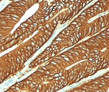
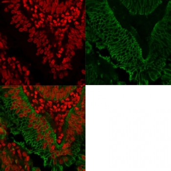
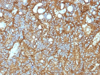
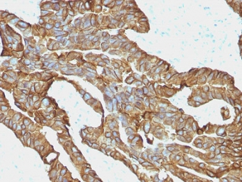
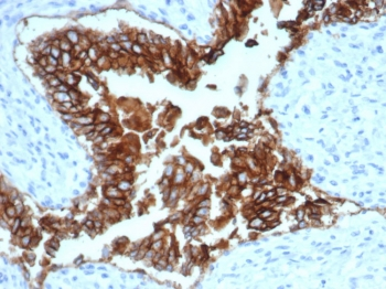
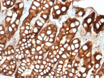
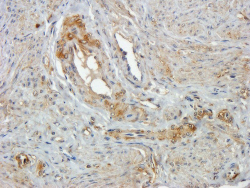
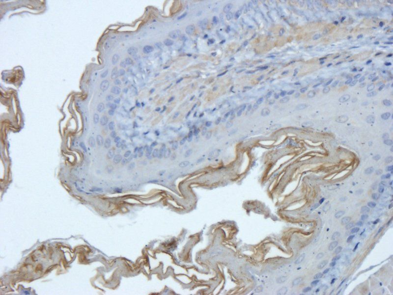
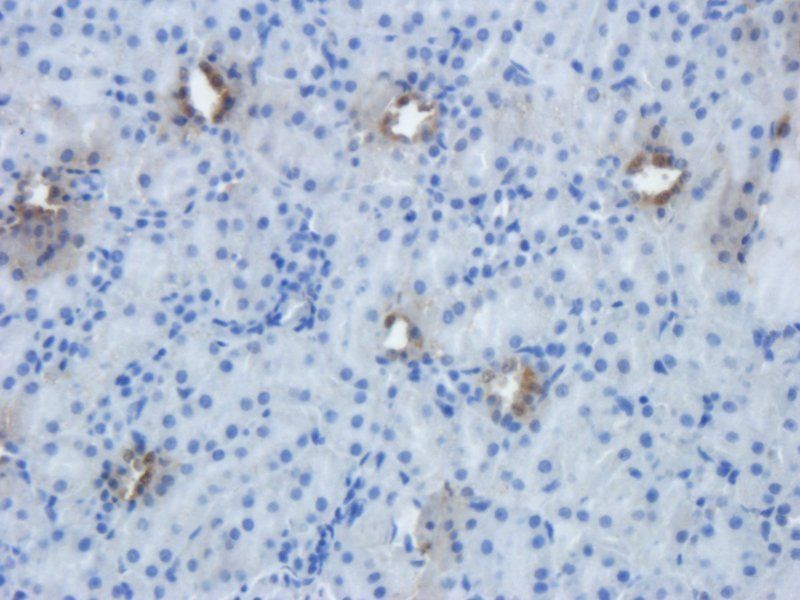
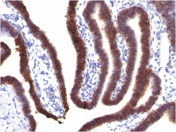
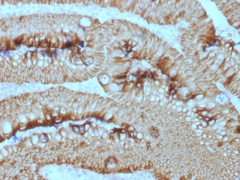
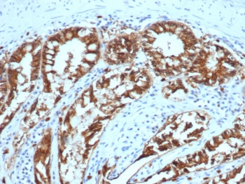
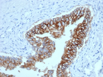
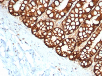
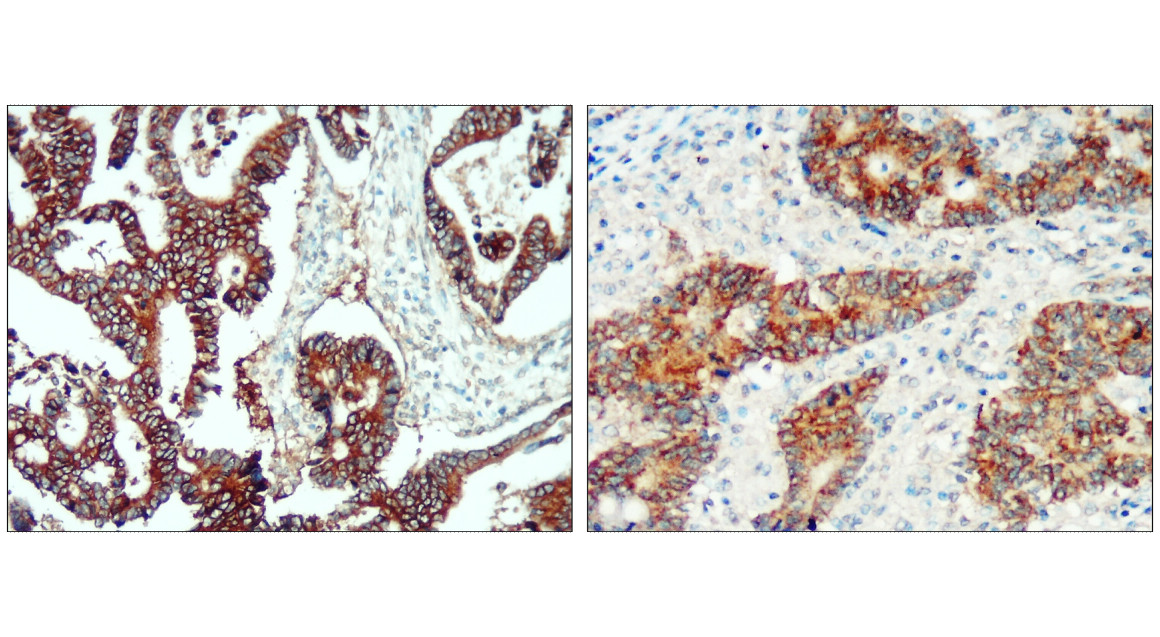
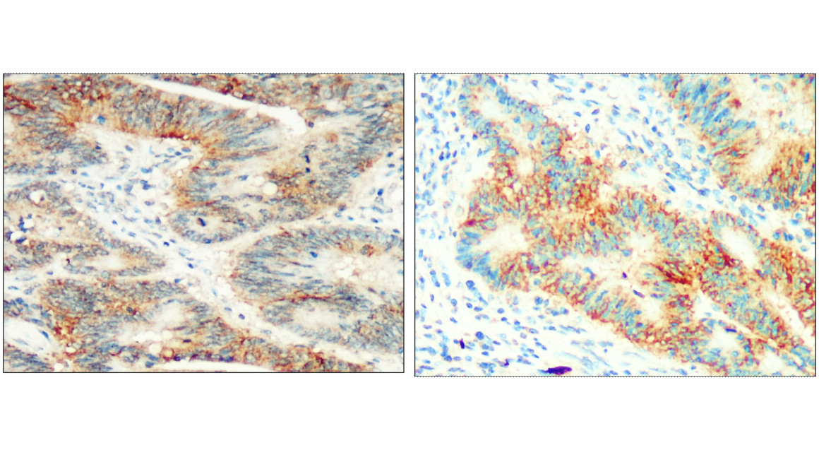
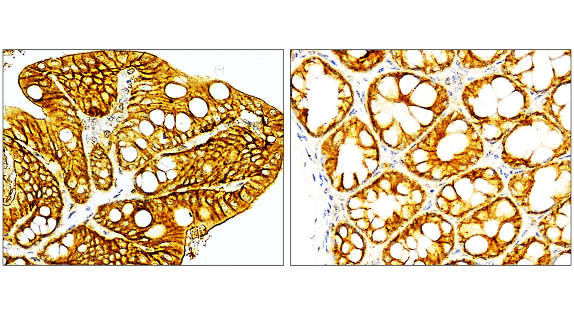
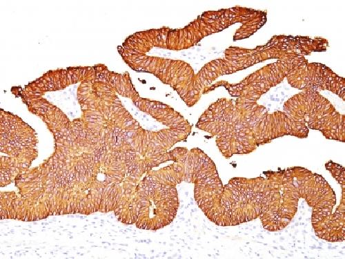
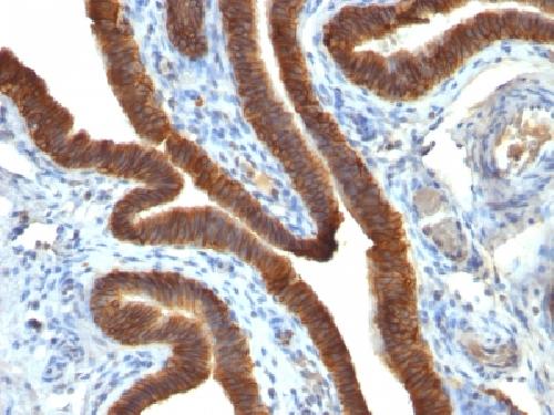
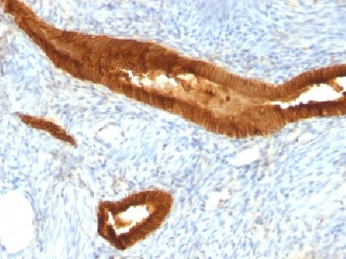
IHC testing of FFPE colon carcinoma stained with EpCAM antibody (clone VU-1D9).
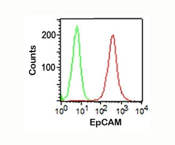
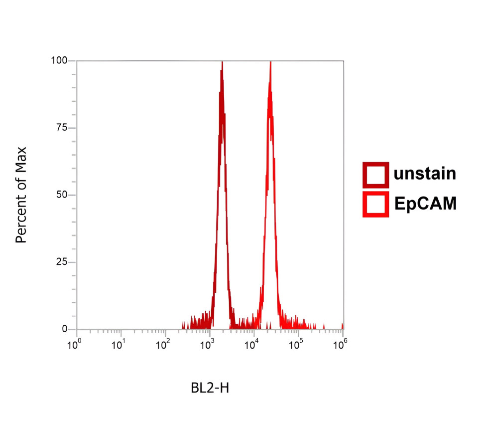
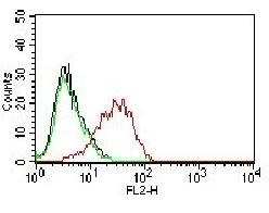
FACS staining (surface) of HT29 cells using EpCAM antibody (VU-1D9, red) and isotype control (green).
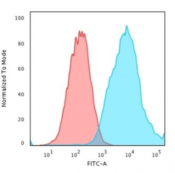
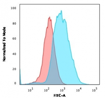
Flow cytometry testing of PFA-fixed human MCF7 cells with EpCAM antibody (clone VU-1D9); Red=isotype control, Blue=CF488-labeled EpCAM antibody.
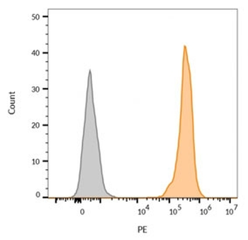
Flow cytometry testing of human MCF7 cells with (orange) and without (gray) CF555-labeled EpCAM antibody (clone VU-1D9).
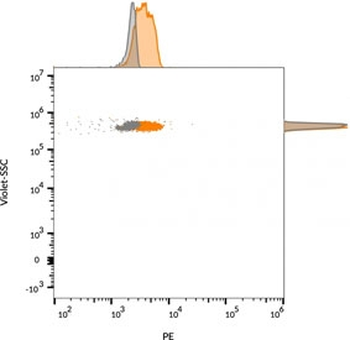
Flow cytometry testing of bead-bound exosomes derived from human MCF-7 cells with (orange) and without (gray) CF555-labeled EpCAM antibody (clone VU-1D9).
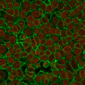
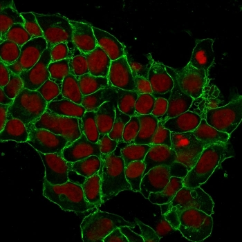
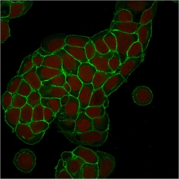
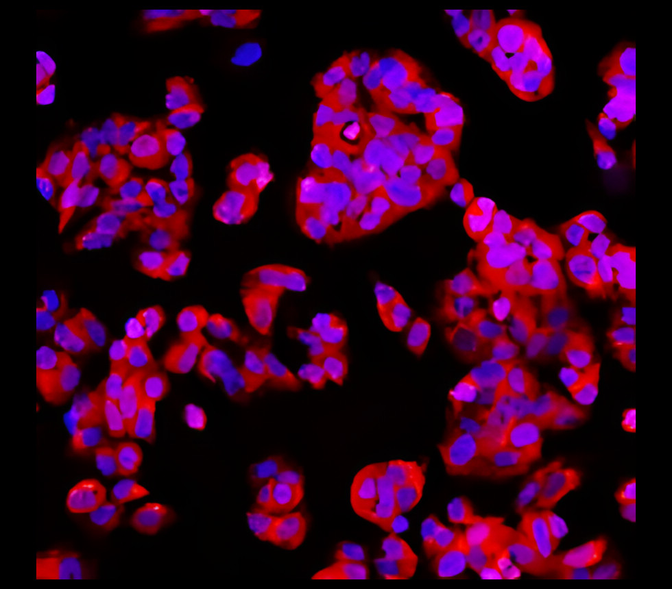
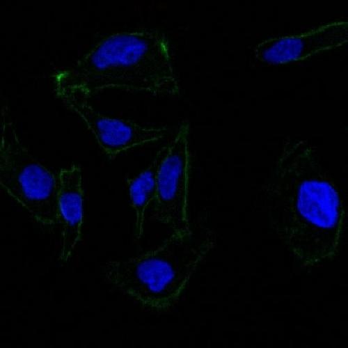
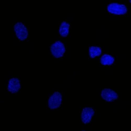
Immunofluorescent staining of human MCF7 cells with EpCAM antibody (clone VU-1D9, green) and Nucspot nuclear stain (red).
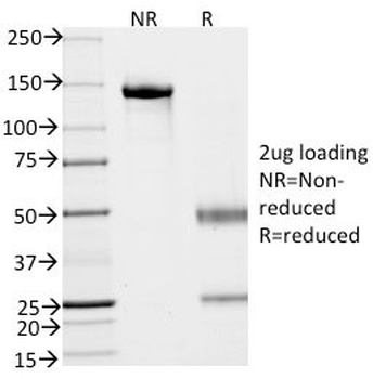
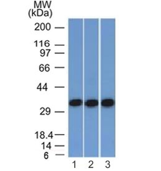
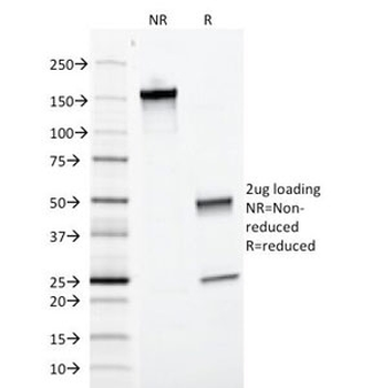
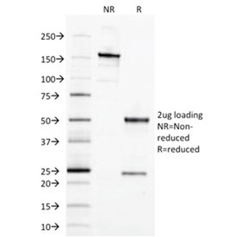
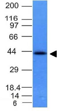
SDS-PAGE analysis of purified, BSA-free EpCAM antibody (clone VU-1D9) as confirmation of integrity and purity.
- Item 1 of 11
EpCAM Antibody / Extracellular domain [orb606357]
FACS, IF, IHC-P, WB
Canine, Feline, Human
Mouse
Monoclonal
Unconjugated
20 μg, 100 μg - Item 1 of 5
- Item 1 of 9
EpCAM Antibody [orb639787]
IF, IHC-P, WB
Canine, Feline, Human
Mouse
Monoclonal
Unconjugated
20 μg, 100 μg - Item 1 of 8
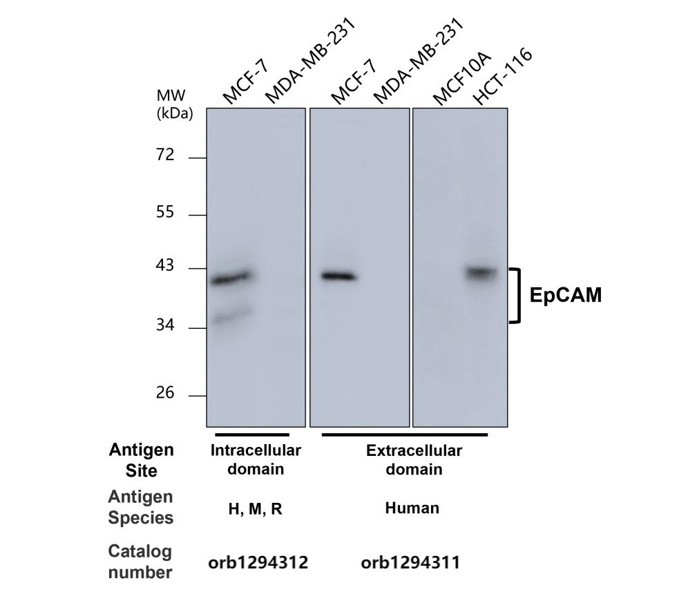
EpCAM (Extracellular domain) antibody [orb1294311]
IF, IHC, WB
Human
Rabbit
Polyclonal
Unconjugated
100 μl, 25 μl - Item 1 of 7
EpCAM antibody [orb389162]
FC, IF, IHC-P, WB
Canine, Feline, Human
Mouse
Monoclonal
Unconjugated
100 μg, 20 μg
Submit a review
Filter by Rating

- 5 stars

- 4 stars
- 3 stars
- 2 stars
- 1 stars
