You have no items in your shopping cart.
Cart summary
Item 1 of 4
Item 1 of 4
ENO1 Antibody (Center)
Catalog Number: orb1788327

| Catalog Number | orb1788327 |
|---|---|
| Category | Antibodies |
| Description | Purified Rabbit Polyclonal Antibody (Pab) |
| Species/Host | Rabbit |
| Clonality | Polyclonal |
| Tested applications | FC, IF, IHC-P, WB |
| Predicted Reactivity | Other, Rat |
| Reactivity | Human, Mouse |
| Isotype | Rabbit IgG |
| Immunogen | 178-205 aa |
| Dilution range | IF: 1:10~50, WB: 1:1000, IHC-P: 1:10~50, FC: 1:10~50 |
| Form/Appearance | Purified polyclonal antibody supplied in PBS with 0.09% (W/V) sodium azide. This antibody is prepared by Saturated Ammonium Sulfate (SAS) precipitation followed by dialysis against PBS. |
| Conjugation | Unconjugated |
| MW | 47169 Da |
| Target | This ENO1 antibody is generated from rabbits immunized with a KLH conjugated synthetic peptide between 178-205 amino acids from the Central region of human ENO1. |
| UniProt ID | P06733 |
| NCBI | NP_001188412.1, NP_001419.1 |
| Storage | Maintain refrigerated at 2-8°C for up to 2 weeks. For long term storage store at -20°C in small aliquots to prevent freeze-thaw cycles |
| Alternative names | ENO1; ENO1L1; MBPB1; MPB1; Alpha-enolase; 2-phosph Read more... |
| Note | For research use only |
| Expiration Date | 12 months from date of receipt. |

ENO1 Antibody (Center) flow cytometric analysis of Hela cells (right histogram) compared to a negative control cell (left histogram). FITC-conjugated donkey-anti-rabbit secondary antibodies were used for the analysis.
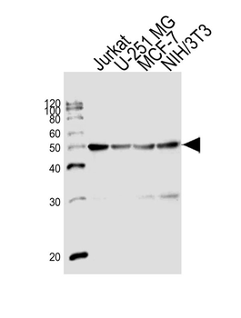
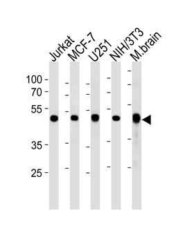
Western blot analysis of lysates from Jurkat, U-251 MG, MCF-7, mouse NIH/3T3 cell line (from left to right), using ENO1 Antibody (Center). Diluted at 1:1000 at each lane. A goat anti-rabbit IgG H&L (HRP) at 1:10000 dilution was used as the secondary antibody. Lysates at 20ug per lane.
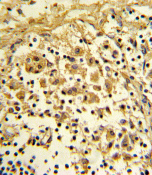
Formalin-fixed and paraffin-embedded human lymph reacted with ENO1 Antibody (Center), which was peroxidase-conjugated to the secondary antibody, followed by DAB staining. This data demonstrates the use of this antibody for immunohistochemistry; clinical relevance has not been evaluated.
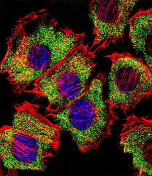
Fluorescent confocal image of C2C12 cell stained with ENO1 Antibody (Center).C2C12 cells were fixed with 4% PFA (20 min), permeabilized with Triton X-100 (0.1%, 10 min), then incubated with ENO1 primary antibody (1:25, 1 h at 37°C). For secondary antibody, Alexa Fluor 488 conjugated donkey anti-rabbit antibody (green) was used (1:400, 50 min at 37°C).Cytoplasmic actin was counterstained with Alexa Fluor 555 (red) conjugated Phalloidin (7units/ml, 1 h at 37°C). Nuclei were counterstained with DAPI (blue) (10 µg/ml, 10 min). ENO1 immunoreactivity is localized to Cytoplasm significantly.
- Item 1 of 4
ENO1 Antibody (Center) [orb1930014]
FC, IF, IHC-P, WB
Other, Rat
Human, Mouse
Rabbit
Polyclonal
Unconjugated
50 μl, 100 μl