You have no items in your shopping cart.
Cart summary
Item 1 of 5
Item 1 of 5
EN1 (Engrailed 1) Antibody (N-term)
Catalog Number: orb1929397

| Catalog Number | orb1929397 |
|---|---|
| Category | Antibodies |
| Description | Affinity Purified Rabbit Polyclonal Antibody (Pab) |
| Species/Host | Rabbit |
| Clonality | Polyclonal |
| Clone Number | RB13885 |
| Tested applications | IF, IHC-P, WB |
| Reactivity | Human, Mouse |
| Isotype | Rabbit IgG |
| Dilution range | IF: 1:10~50, IF: 1:25, WB: 1:2000, IHC-P: 1:10~50, IHC-P: 1:25 |
| Form/Appearance | Purified polyclonal antibody supplied in PBS with 0.09% (W/V) sodium azide. This antibody is purified through a protein A column, followed by peptide affinity purification. |
| Conjugation | Unconjugated |
| MW | 40115 Da |
| Target | This EN1 (Engrailed 1) antibody is generated from rabbits immunized with a KLH conjugated synthetic peptide between 1-30 amino acids from the N-terminal region of human EN1 (Engrailed 1). |
| UniProt ID | Q05925 |
| NCBI | NP_001417.3 |
| Storage | Maintain refrigerated at 2-8°C for up to 2 weeks. For long term storage store at -20°C in small aliquots to prevent freeze-thaw cycles |
| Alternative names | Homeobox protein engrailed-1, Homeobox protein en- Read more... |
| Note | For research use only |
| Expiration Date | 12 months from date of receipt. |

Staining EN1 in Human tonsil tissue sections by Immunohistochemistry (IHC-P - paraformaldehyde-fixed, paraffin-embedded sections). Tissue was fixed with formaldehyde and blocked with 3% BSA for 0.5 hour at room temperature; antigen retrieval was by heat mediation with a citrate buffer (pH6). Samples were incubated with primary antibody (1/25) for 1 hours at 37°C. A undiluted biotinylated goat polyvalent antibody was used as the secondary antibody.
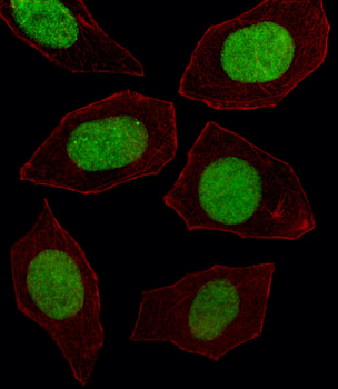
Immunofluorescent analysis of 4% paraformaldehyde-fixed, 0.1% Triton X-100 permeabilized U-2 OS (Human Sarcoma cell line) cells labeling HME1 at 1/25 dilution, followed by Alexa Fluor 488-conjugated goat anti-rabbit IgG secondary antibody at 1/400 dilution (green). Immunofluorescence image showing nucleus and nucleoli staining on U-2 OS cell line. Cytoplasmic actin is detected with Alexa Fluor 555 conjugated with Phalloidin at 1/100 dilution (red).
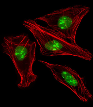
Fluorescent image of U251 cell stained with EN1 Antibody (N-term).U251 cells were fixed with 4% PFA (20 min), permeabilized with Triton X-100 (0.1%, 10 min), then incubated with EN1 primary antibody (1:25, 1 h at 37°C). For secondary antibody, Alexa Fluor 488 conjugated donkey anti-rabbit antibody (green) was used (1:400, 50 min at 37°C).Cytoplasmic actin was counterstained with Alexa Fluor 555 (red) conjugated Phalloidin (7units/ml, 1 h at 37°C).EN1 immunoreactivity is localized to Nucleus significantly.

Formalin-fixed and paraffin-embedded human lung carcinoma tissue reacted with EN1 antibody (N-term), which was peroxidase-conjugated to the secondary antibody, followed by DAB staining. This data demonstrates the use of this antibody for immunohistochemistry; clinical relevance has not been evaluated.
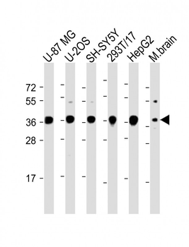
All lanes: Anti-EN1 (Engrailed 1) Antibody (N-term) at 1:2000 dilution. Lane 1: U-87 MG whole cell lysate. Lane 2: U-2OS whole cell lysate. Lane 3: SH-SY5Y whole cell lysate. Lane 4: 293T/17 whole cell lysate. Lane 5: HepG2 whole cell lysate. Lane 6: Mouse brain lysate. Lysates/proteins at 20 µg per lane. Secondary Goat Anti-Rabbit IgG, (H+L), Peroxidase conjugated at 1/10000 dilution. Predicted band size: 40 kDa. Blocking/Dilution buffer: 5% NFDM/TBST.

EN1 (Engrailed 1) Antibody (N-term) [orb1166388]
IF, IHC-P, WB
Human
Rabbit
Polyclonal
Unconjugated
100 μl, 30 μl
