You have no items in your shopping cart.
Cart summary
Item 1 of 7
Item 1 of 7
ELAVL1 Antibody
Catalog Number: orb1928182

| Catalog Number | orb1928182 |
|---|---|
| Category | Antibodies |
| Description | Affinity Purified Rabbit Polyclonal Antibody (Pab) |
| Species/Host | Rabbit |
| Clonality | Polyclonal |
| Clone Number | RB22938 |
| Tested applications | FC, IHC-P, WB |
| Reactivity | Human, Mouse |
| Isotype | Rabbit IgG |
| Dilution range | WB: 1:1000, WB: 1:2000, IHC-P: 1:50~100, IHC-P: 1:100, IHC-P: 1:25, FC: 1:25, FC: 1:25 |
| Form/Appearance | Purified polyclonal antibody supplied in PBS with 0.09% (W/V) sodium azide. This antibody is purified through a protein A column, followed by peptide affinity purification. |
| Conjugation | Unconjugated |
| MW | 36092 Da |
| Target | This ELAVL1 antibody is generated from rabbits immunized with human ELAVL1 recombinant protein. |
| UniProt ID | Q15717 |
| NCBI | NP_001410.2 |
| Storage | Maintain refrigerated at 2-8°C for up to 2 weeks. For long term storage store at -20°C in small aliquots to prevent freeze-thaw cycles |
| Alternative names | ELAV-like protein 1, Hu-antigen R, HuR, ELAVL1, HU Read more... |
| Note | For research use only |
| Expiration Date | 12 months from date of receipt. |

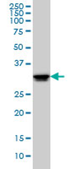
Western blot analysis of ELAVL1 (arrow) using rabbit polyclonal ELAVL1 Antibody. 293 cell lysates (2 ug/lane) either nontransfected (Lane 1) or transiently transfected (Lane 2) with the ELAVL1 gene.

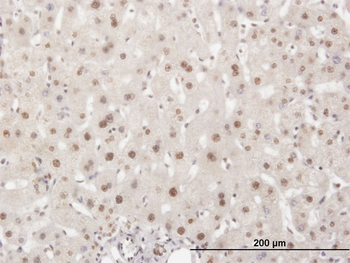
Formalin-fixed and paraffin-embedded human brain tissue reacted with ELAVL1 Antibody, which was peroxidase-conjugated to the secondary antibody, followed by DAB staining. This data demonstrates the use of this antibody for immunohistochemistry; clinical relevance has not been evaluated.
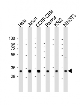
All lanes: Anti-ELAVL1 Antibody at 1:2000 dilution. Lane 1: Hela whole cell lysate. Lane 2: Jurkat whole cell lysate. Lane 3: CCRF-CEM whole cell lysate. Lane 4: Ramos whole cell lysate. Lane 5: K562 whole cell lysate. Lane 6: NIH/3T3 whole cell lysate. Lysates/proteins at 20 µg per lane. Secondary Goat Anti-Rabbit IgG, (H+L), Peroxidase conjugated at 1/10000 dilution. Predicted band size: 36 kDa. Blocking/Dilution buffer: 5% NFDM/TBST.
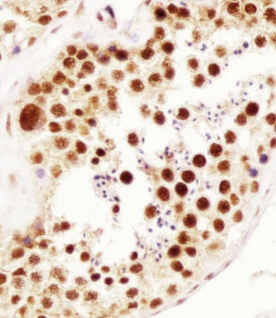
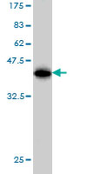
Staining ELAVL1 in human testis tissue sections by Immunohistochemistry (IHC-P - paraformaldehyde-fixed, paraffin-embedded sections). Tissue was fixed with formaldehyde and blocked with 3% BSA for 0.5 hour at room temperature; antigen retrieval was by heat mediation with a citrate buffer (pH6). Samples were incubated with primary antibody (1/25) for 1 hours at 37°C. A undiluted biotinylated goat polyvalent antibody was used as the secondary antibody.
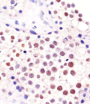
Staining ELAVL1 in human testis tissue sections by Immunohistochemistry (IHC-P - paraformaldehyde-fixed, paraffin-embedded sections). Tissue was fixed with formaldehyde and blocked with 3% BSA for 0.5 hour at room temperature; antigen retrieval was by heat mediation with a citrate buffer (pH6). Samples were incubated with primary antibody (1/100) for 1 hours at 37°C. A undiluted biotinylated goat polyvalent antibody was used as the secondary antibody.
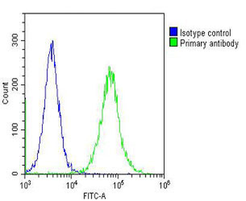
Overlay histogram showing Hela cells stained (green line). The cells were fixed with 2% paraformaldehyde (10 min) and then permeabilized with 90% methanol for 10 min. The cells were then icubated in 2% bovine serum albumin to block non-specific protein-protein interactions followed by the antibody (1:25 dilution) for 60 min at 37°C. The secondary antibody used was Goat-Anti-Rabbit IgG, DyLight 488 Conjugated Highly Cross-Adsorbed at 1/200 dilution for 40 min at 37°C. Isotype control antibody (blue line) was rabbit IgG (1 μg/1x10^6 cells) used under the same conditions. Acquisition of > 10000 events was performed.
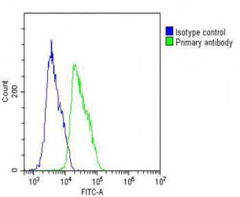
Overlay histogram showing MCF-7 cells stained (green line). The cells were fixed with 2% paraformaldehyde (10 min) and then permeabilized with 90% methanol for 10 min. The cells were then icubated in 2% bovine serum albumin to block non-specific protein-protein interactions followed by the antibody (1:25 dilution) for 60 min at 37°C. The secondary antibody used was Goat-Anti-Rabbit IgG, DyLight 488 Conjugated Highly Cross-Adsorbed at 1/200 dilution for 40 min at 37°C. Isotype control antibody (blue line) was rabbit IgG (1 μg/1x10^6 cells) used under the same conditions. Acquisition of > 10000 events was performed.
- Item 1 of 8
HuR / ELAVL1 Antibody, KO Validated [orb1274490]
IF, IHC, WB
Human, Mouse, Rat
Rabbit
Polyclonal
Unconjugated
100 μl - Item 1 of 7
- Item 1 of 5
ELAVL1 monoclonal antibody (M02), clone 4G8 [orb2293258]
ELISA, IF, IHC-P, WB
Human
Mouse
Monoclonal
Unconjugated
100 μg - Item 1 of 6
HuR / ELAVL1 Antibody, KO Validated [orb1274439]
IF, IHC, WB
Human, Mouse
Rabbit
Polyclonal
Unconjugated
100 μl - Item 1 of 3