You have no items in your shopping cart.
Cart summary
Item 1 of 5
Item 1 of 5
Eif2ak3 antibody
Catalog Number: orb750495

| Catalog Number | orb750495 |
|---|---|
| Category | Antibodies |
| Description | Eif2ak3 antibody |
| Species/Host | Rabbit |
| Clonality | Polyclonal |
| Tested applications | ELISA, IF, IHC, IP, WB |
| Reactivity | Mouse |
| Isotype | Antiserum |
| Immunogen | This whole rabbit serum was prepared by repeated immunizations with a recombinant fusion protein from amino acids 601-1115 of mouse deltaN PERK. |
| Concentration | 80 mg/mL |
| Dilution range | ELISA: 1:4,000 - 1:20,000, IHC: 1:1,000, IF: User Optimized, IP: 10-30ul, WB: 1:500 – 1:3000 |
| Form/Appearance | Liquid (sterile filtered) |
| Purity | This antiserum is directed against PERK and reacts with the PERK from mouse tissues. Reactivity to other species is unknown. |
| Conjugation | Unconjugated |
| UniProt ID | Q9Z2B5 |
| NCBI | 32451785 |
| Storage | Store vial at -20° C prior to opening. Aliquot contents and freeze at -20° C or below for extended storage. Avoid cycles of freezing and thawing. Centrifuge product if not completely clear after standing at room temperature. This product is stable for several weeks at 4° C as an undiluted liquid. Dilute only prior to immediate use. |
| Buffer/Preservatives | 0.01% (w/v) Sodium Azide |
| Alternative names | rabbit anti-PKR-like Endoplasmic Reticulum Kinase Read more... |
| Note | For research use only |
| Application notes | This antiserum has been tested for use in western blotting, immunoprecipitation and immunohistochemistry. Specific conditions for reactivity should be optimized by the end user. Expect bands approximately 150kDa by western blotting in the appropriate cell lysate or extract. |
| Expiration Date | 12 months from date of receipt. |
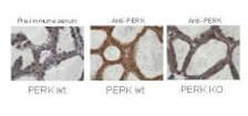
Immunohistochemistry staining of mouse mammary gland samples from lactating mice (L10) with Biorbyt's anti-PERK. Positive staining signal observed in wild type mouse sample with anti-PERK staining only (middle image), but not in the knock out mouse sample (right image) and pre-immune serum staining (left image) The anti-PERK was diluted 1:1000 in 5% goat serum in PBS and allowed to incubate for 2h at room temperature in a humidified chamber.
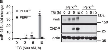
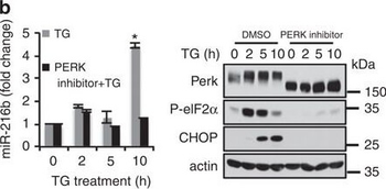
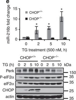
PERK-dependent miR-216b induction. (a) PERK+ /+ and PERK−/− MEFs were treated with 500 nM TG for indicated times. MiR-216b was assessed by qPCR (left graph); PERK and CHOP were assessed by immunoblot (right). (b) MiR-216b levels were quantified by qPCR following exposure of cells to thapsigargin and a small-molecule PERK inhibitor (left). PERK, eIF2α-p and CHOP induction were assessed by immunoblot (right). (c–e) MEFs of the indicated genotype were treated with TG (500 nM) for indicated intervals. Protein extracts from these cells were immunoblotted for the proteins as indicated (lower panels) and miR-216b levels were quantified by qPCR (upper panels; n=3). (f) CHOP−/− MEFs were transfected with vector or CHOP and 2 days later treated with TG (500 nM) for indicated intervals. Protein extracts from these cells were immunoblotted for CHOP and miR-216b levels quantified by qPCR (n=3). (g) MiR-216b expression and (h) c-Jun mRNA levels were analysed in MMTV-Neu tumours from either PERK+ /+ or a PERK−/− background. Data represent mean±s.d. of three independent observations. Statistical significance was analysed analysed by t-test. (*P < 0.05, WT versus −/−).
PERK-dependent miR-216b induction. (a) PERK+ /+ and PERK−/− MEFs were treated with 500 nM TG for indicated times. MiR-216b was assessed by qPCR (left graph); PERK and CHOP were assessed by immunoblot (right). (b) MiR-216b levels were quantified by qPCR following exposure of cells to thapsigargin and a small-molecule PERK inhibitor (left). PERK, eIF2α-p and CHOP induction were assessed by immunoblot (right). (c–e) MEFs of the indicated genotype were treated with TG (500 nM) for indicated intervals. Protein extracts from these cells were immunoblotted for the proteins as indicated (lower panels) and miR-216b levels were quantified by qPCR (upper panels; n=3). (f) CHOP−/− MEFs were transfected with vector or CHOP and 2 days later treated with TG (500 nM) for indicated intervals. Protein extracts from these cells were immunoblotted for CHOP and miR-216b levels quantified by qPCR (n=3). (g) MiR-216b expression and (h) c-Jun mRNA levels were analysed in MMTV-Neu tumours from either PERK+ /+ or a PERK−/− background. Data represent mean±s.d. of three independent observations. Statistical significance was analysed analysed by t-test. (*P < 0.05, WT versus −/−).
PERK-dependent miR-216b induction. (a) PERK+ /+ and PERK−/− MEFs were treated with 500 nM TG for indicated times. MiR-216b was assessed by qPCR (left graph); PERK and CHOP were assessed by immunoblot (right). (b) MiR-216b levels were quantified by qPCR following exposure of cells to thapsigargin and a small-molecule PERK inhibitor (left). PERK, eIF2α-p and CHOP induction were assessed by immunoblot (right). (c–e) MEFs of the indicated genotype were treated with TG (500 nM) for indicated intervals. Protein extracts from these cells were immunoblotted for the proteins as indicated (lower panels) and miR-216b levels were quantified by qPCR (upper panels; n=3). (f) CHOP−/− MEFs were transfected with vector or CHOP and 2 days later treated with TG (500 nM) for indicated intervals. Protein extracts from these cells were immunoblotted for CHOP and miR-216b levels quantified by qPCR (n=3). (g) MiR-216b expression and (h) c-Jun mRNA levels were analysed in MMTV-Neu tumours from either PERK+ /+ or a PERK−/− background. Data represent mean±s.d. of three independent observations. Statistical significance was analysed analysed by t-test. (*P < 0.05, WT versus −/−).
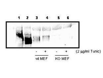
Western blot analysis using Biorbyt's anti-PERK to detect PERK in cell lysates. 300 µg PERK over-expressing 293T cell lysate (lanes 1 & 2), or 800 ug wild type (Lanes 3 & 4), and PERK knock out (lanes 5 & 6) MEF cell lysate were immunoprecipated with 15 µL anti-PERK, followed by western blotting with 1:1000 dilution of anti-PERK in 5% milk/TBST buffer. Lane 1, 293T cells over-expressing Myc-PERK wt, Lane 2, 293T cells over-expressing Myc-PERK K618A.
- Item 1 of 6
PERK/EIF2AK3 Antibody [orb1294328]
IF, IHC, WB
Human, Mouse
Rabbit
Polyclonal
Unconjugated
100 μl, 25 μl - Item 1 of 4
- Item 1 of 4
PERK antibody [orb770922]
ELISA, IF, IHC-P, WB
Human, Mouse, Rat
Rabbit
Polyclonal
Unconjugated
50ul, 100ul - Item 1 of 2
- Item 1 of 3


Submit a review
Filter by Rating

- 5 stars

- 4 stars
- 3 stars
- 2 stars
- 1 stars
