You have no items in your shopping cart.
Cart summary
Item 1 of 2
Item 1 of 2
E2F-1 (phospho-S364) antibody
Catalog Number: orb345473

| Catalog Number | orb345473 |
|---|---|
| Category | Antibodies |
| Description | E2F-1 (phospho-S364) antibody |
| Species/Host | Rabbit |
| Clonality | Polyclonal |
| Tested applications | ELISA, IHC, WB |
| Reactivity | Human, Mouse |
| Isotype | IgG |
| Immunogen | This affinity purified antibody was prepared from whole rabbit serum produced by repeated immunizations with a synthetic peptide corresponding to an internal region near amino acids 350-375 of Human E2F-1. |
| Concentration | 1.0 mg/mL |
| Dilution range | ELISA: 1:20,000 - 1:100,000, IHC: 2 mg/ml - 20 µg/ml, WB: 1:250 - 1:2,000 |
| Form/Appearance | Liquid (sterile filtered) |
| Purity | This affinity purified antibody is directed against the phosphorylated form of human E2F-1 at the pS364 residue. The product was affinity purified from monospecific antiserum by immunoaffinity purification. Antiserum was first purified against the phosphorylated form of the immunizing peptide. The resultant affinity purified antibody was then cross adsorbed against the non-phosphorylated form of the immunizing peptide. Reactivity occurs against human E2F-1 pS364 protein and the antibody is specific for the phosphorylated form of the protein. Reactivity with non-phosphorylated human E2F-1 is minimal by ELISA. The antibody does not cross-react with E2F-1 phosphorylated at other sites. A BLAST analysis was used to suggest reactivity with this protein from human and chimpanzee based on 100% homology for the immunogen sequence. Cross reactivity with E2F-1 homologues from other sources has not been determined. |
| Conjugation | Unconjugated |
| UniProt ID | Q01094 |
| NCBI | 12669911 |
| Storage | Store vial at -20° C prior to opening. Aliquot contents and freeze at -20° C or below for extended storage. Avoid cycles of freezing and thawing. Centrifuge product if not completely clear after standing at room temperature. This product is stable for several weeks at 4° C as an undiluted liquid. Dilute only prior to immediate use. |
| Buffer/Preservatives | 0.01% (w/v) Sodium Azide |
| Alternative names | rabbit anti-E2F-1 pS364 Antibody, E2F 1 antibody, Read more... |
| Note | For research use only |
| Application notes | This affinity purified antibody has been tested for use in ELISA, immunohistochemistry and by western blot. Specific conditions for reactivity should be optimized by the end user. Expect a band approximately 47 kDa in size corresponding to phosphorylated E2F-1 by western blotting in the appropriate cell lysate or extract. Less than 0.5% reactivity is observed against the non-phosphorylated form of the immunizing peptide. This antibody is phospho specific for pS364 of E2F-1. |
| Expiration Date | 12 months from date of receipt. |
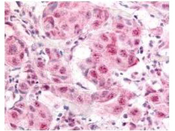
Biorbyt's Affinity Purified anti-E2F-1 pS364 antibody was used at a 10 µg/ml to detect nuclear and occasionally cytoplasmic signal in a variety of tissues including multi-human, multi-brain and multi-cancer slides. Within the multi-tumor block, the antibody showed variable levels of nuclear staining between individual tumors, with some tumors showing strong staining. This image shows E2F-1 pS364 staining of human breast carcinoma. Tissue was formalin-fixed and paraffin embedded.
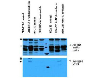
Western blot using Biorbyt's Affinity Purified anti-E2F-1 pS364 antibody shows detection of a band at ~47 kDa corresponding to phosphorylated E2F-1 in induced cell lysates. Panel A shows reactivity using a control antibody reactive to all forms of E2F (arrowheads). Panel B shows specific reactivity against phosphorylated E2F-1 (arrowheads) using our anti-E2F-1 pS364 antibody. Lysates are as follows: CRE/E2F-1 are CRE cells derived from mouse NIH3T3 line transfected with human E2F-1, NIH-3T3 used as a negative control, and MDA-MB-231 cells are a human breast cancer line. As indicated each lysate was prepared from untreated cells and cells treated with 2 µM Doxorubicin used as a DNA damaging agent. In addition the MDA-MB-231 cells were also treated with genistein, a mild DNA damaging agent. The figure shows the same membrane first probed with the anti-E2F-1 pS364 antibody used at a 1:250 dilution, then stripped and re-probed with the pan E2F antibody used as a positive control. The positive control antibody clearly shows an E2F-1 band in all human cell lines, but not mouse cells. Treatment with doxorubicin increases the expression of E2F-1 as shown in Panel A. After film development, images were overlapped to confirm that specific anti-E2F-1 pS364 staining shown treated human cells in Panel B specifically aligns with E2F-1 staining shown in Panel A. Blots can be processed with HRP conjugated Gt-a-Rabbit IgG MX10 orb347654 for 45 min at room temperature for ECL detection.
- Item 1 of 5
E2F1 (phospho-Ser364) antibody [orb5095]
FC, ICC, IHC-P, WB
Mouse, Rat
Human
Rabbit
Polyclonal
Unconjugated
200 μl, 50 μl, 100 μl 
phospho-E2F1 (Ser364) Antibody (BF750) [orb1582277]
FC, ICC, IF
Mouse, Rat
Human
Rabbit
Polyclonal
BF750
100 μlphospho-E2F1 (Ser364) Antibody (BF680) [orb1582278]
FC, ICC, IF
Mouse, Rat
Human
Rabbit
Polyclonal
BF680
100 μlphospho-E2F1 (Ser364) Antibody (BF647) [orb1582279]
FC, ICC, IF
Mouse, Rat
Human
Rabbit
Polyclonal
BF647
100 μl


Submit a review
Filter by Rating

- 5 stars

- 4 stars
- 3 stars
- 2 stars
- 1 stars
