You have no items in your shopping cart.
Cart summary
Item 1 of 7
Item 1 of 7
DVL1 Antibody (Center)
Catalog Number: orb1937565

| Catalog Number | orb1937565 |
|---|---|
| Category | Antibodies |
| Description | Affinity Purified Rabbit Polyclonal Antibody (Pab) |
| Species/Host | Rabbit |
| Clonality | Polyclonal |
| Clone Number | RB31716 |
| Tested applications | IF, IHC-P, WB |
| Predicted Reactivity | Rat |
| Reactivity | Human, Mouse |
| Isotype | Rabbit IgG |
| Antibody Type | Primary Antibody |
| Dilution range | IF: 1:10~50, IF: 1:25, WB: 1:1000, WB: 1:2000, WB: 1:1000, IHC-P: 1:10~50, IHC-P: 1:25 |
| Form/Appearance | Purified polyclonal antibody supplied in PBS with 0.09% (W/V) sodium azide. This antibody is purified through a protein A column, followed by peptide affinity purification. |
| Conjugation | Unconjugated |
| MW | 75187 Da |
| Target | This DVL1 antibody is generated from rabbits immunized with a KLH conjugated synthetic peptide between 442-470 amino acids from the Central region of human DVL1. |
| UniProt ID | O14640 |
| NCBI | NP_004412.2 |
| Storage | Maintain refrigerated at 2-8°C for up to 2 weeks. For long term storage store at -20°C in small aliquots to prevent freeze-thaw cycles |
| Alternative names | Segment polarity protein dishevelled homolog DVL-1 Read more... |
| Note | For research use only |
| Expiration Date | 12 months from date of receipt. |

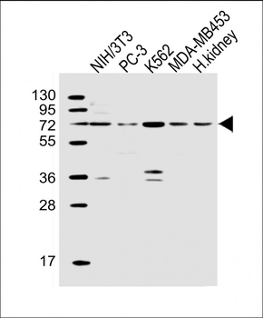
All lanes: Anti-DVL1 Antibody (Center) at 1:2000 dilution. Lane 1: K562 whole cell lysates. Lane 2: MDA-MB-453 whole cell lysates. Lane 3: PC-3 whole cell lysates. Lane 4: human kidney lysates.Lysates/proteins at 20 µg per lane. Secondary Goat Anti-Rabbit IgG, (H+L), Peroxidase conjugated at 1/10000 dilution. Predicted band size: 75 kDa. Blocking/Dilution buffer: 5% NFDM/TBST.
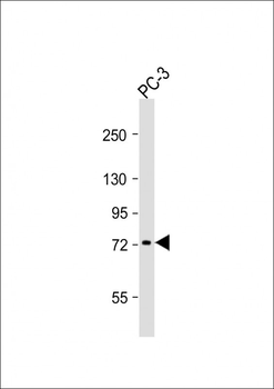
Anti-DVL1 Antibody (Center) at 1:1000 dilution + PC-3 whole cell lysates.Lysates/proteins at 20 µg per lane. Secondary Goat Anti-Rabbit IgG, (H+L), Peroxidase conjugated at 1/10000 dilution. Predicted band size: 75 kDa. Blocking/Dilution buffer: 5% NFDM/TBST.
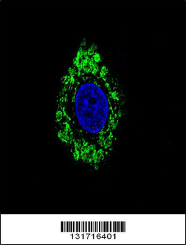
Confocal immunofluorescent analysis of DVL1 Antibody (Center) with HepG2 cell followed by Alexa Fluor 488-conjugated goat anti-rabbit lgG (green). DAPI was used to stain the cell nuclear (blue).
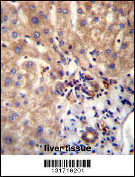
DVL1 Antibdy (Center) immunohistochemistry analysis in formalin fixed and paraffin embedded human liver tissue followed by peroxidase conjugation of the secondary antibody and DAB staining. This data demonstrates the use of DVL1 Antibdy (Center) for immunohistochemistry. Clinical relevance has not been evaluated.
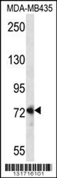
DVL1 Antibody (Center) western blot analysis in MDA-MB435 cell line lysates (35 ug/lane). This demonstrates the DVL1 antibody detected the DVL1 protein (arrow).
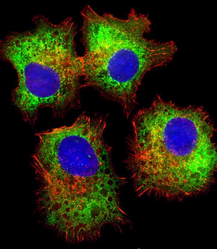
Immunofluorescent analysis of 4% paraformaldehyde-fixed, 0.1% Triton X-100 permeabilized HepG2 (human liver hepatocellular carcinoma cell line) cells labeling DVL1 at 1/25 dilution, followed by Dylight 488-conjugated goat anti-rabbit IgG secondary antibody at 1/200 dilution (green). Immunofluorescence image showing cytoplasm staining on HepG2 cell line. Cytoplasmic actin is detected with Dylight 554 Phalloidin at 1/100 dilution (red).The nuclear counter stain is DAPI (blue).
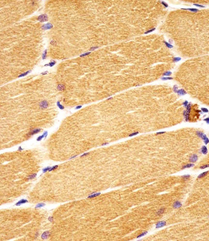
Staining DVL1 in human skeletal muscle sections by Immunohistochemistry (IHC-P - paraformaldehyde-fixed, paraffin-embedded sections). Tissue was fixed with formaldehyde and blocked with 3% BSA for 0.5 hour at room temperature; antigen retrieval was by heat mediation with a citrate buffer (pH6). Samples were incubated with primary antibody (1/25) for 1 hours at 37°C. A undiluted biotinylated goat polyvalent antibody was used as the secondary Antibody.
- Item 1 of 2