You have no items in your shopping cart.
Cart summary
Item 1 of 15
Item 1 of 15
DR5 Antibody
Catalog Number: orb1239348

| Catalog Number | orb1239348 |
|---|---|
| Category | Antibodies |
| Description | DR5 Antibody |
| Species/Host | Rabbit |
| Clonality | Polyclonal |
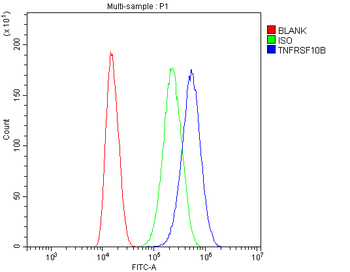
| Tested applications | ELISA, IF, IHC-P, WB |
| Reactivity | Human, Mouse, Rat |
| Isotype | IgG |
| Immunogen | DR5 antibody was raised against a peptide corresponding to 20 amino acids near the carboxy terminus of human DR5 precursor.The immunogen is located within the last 50 amino acids of DR5. |
| Concentration | 1 mg/ml |
| Form/Appearance | Liquid |
| Conjugation | Unconjugated |
| MW | Predicted: 48kD and 45kDObserved: 48kD and 45kD |
| Target | TNFRSF10B |
| UniProt ID | O14763 |
| NCBI | AF012535 |
| Storage | Maintain refrigerated at 2-8°C for up to 2 weeks. For long term storage store at -20°C in small aliquots to prevent freeze-thaw cycles. |
| Buffer/Preservatives | DR5 Antibody is supplied in PBS containing 0.02% sodium azide. |
| Alternative names | DR5 Antibody: DR5, CD262, KILLER, TRICK2, TRICKB, Read more... |
| Note | For research use only |
| Application notes | WB: 0.5-2 μg/mL; IHC-P: 5 μg/mL; IF: 5-10 μg/mL.Antibody validated: Western Blot in human, mouse and rat samples; Immunohistochemistry in mouse samples; Immunocytochemistry in human samples and Immunofluorescence in human, mouse and rat samples. All other applications and species not yet tested. |
| Expiration Date | 12 months from date of receipt. |
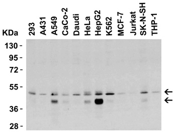
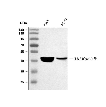
Western Blot Validation in Human Cell Lines. Loading: 15 µg of lysates per lane. Antibodies: DR5 orb1239348, (0.5 µg/mL), 1h incubation at RT in 5% NFDM/TBST. Secondary: Goat anti-rabbit IgG HRP conjugate at 1:10000 dilution.
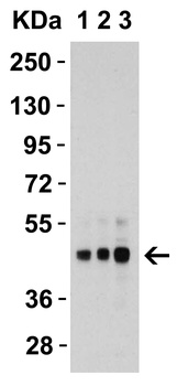
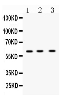
Western Blot Validation in Human HepG2 Cells. Loading: 15 µg of lysates per lane. Antibodies: DR5 orb1239348, 1h incubation at RT in 5% NFDM/TBST. Secondary: Goat anti-rabbit IgG HRP conjugate at 1:10000 dilution. Lane 1: 1 µg/mL, Lane 2: 2 µg/mL, Lane 3: 4 µg/mL.

Western Blot Validation in Mouse and Rat Cell Lines. Loading: 15 µg of lysates per lane. Antibodies: DR5 orb1239348, (2 µg/mL), 1h incubation at RT in 5% NFDM/TBST. Secondary: Goat anti-rabbit IgG HRP.

Western Blot Validation in Mouse Cell Lines. Loading: 15 µg of lysates per lane. Antibodies: DR5 orb1239348, (1 µg/mL), 1h incubation at RT in 5% NFDM/TBST. Secondary: Goat anti-rabbit IgG HRP conjugate at 1:10000 dilution.

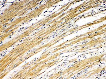
Western Blot Validation in Mouse Heart. Loading: 15 µg of lysatesper lane. Antibodies: DR5 orb1239348, (1 µg/mL), 1h incubation at RT in 5% NFDM/TBST. Secondary: Goat anti-rabbit IgG HRP conjugate at 1:10000 dilution.

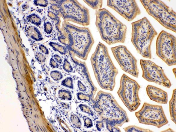
Western Blot Validation in Rat Skeletal Muscle. Loading: 15 µg of lysate per lane. Antibodies: DR5 orb1239348, (1 µg/mL), 1h incubation at RT in 5% NFDM/TBST. Secondary: Goat anti-rabbit IgG HRP conjugate at 1:10000 dilution.
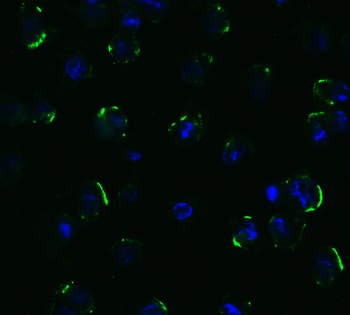
Immunofluorescence Validation of DR5 in Human HepG2 Cells. Immunofluorescent analysis of 4% paraformaldehyde-fixed human HepG2 cells labeling DR5 with orb1239348 at 5 µg/mL, followed by goat anti-rabbit IgG secondary antibody at 1/500 dilution (green) and DAPI (blue).

Immunofluorescence Validation of DR5 in Human Testis. Immunofluorescent analysis of 4% paraformaldehyde-fixed human testis tissue labeling DR5 with orb1239348 at 10 µg/mL, followed by goat anti-rabbit IgG secondary antibody at 1/500 dilution (green) and DAPI (blue).

Immunofluorescence Validation of DR5 in Mouse Pancreas. Immunofluorescent analysis of 4% paraformaldehyde-fixed mouse pancreas tissue labeling DR5 with orb1239348 at 10 µg/mL, followed by goat anti-rabbit IgG secondary antibody at 1/500 dilution (green) and DAPI (blue).

Immunofluorescence Validation of DR5 in Rat Brain. Immunofluorescent analysis of 4% paraformaldehyde-fixed rat brain tissue labeling DR5 with orb1239348 at 5 µg/mL, followed by goat anti-rabbit IgG secondary antibody at 1/500 dilution (green) and DAPI (blue).

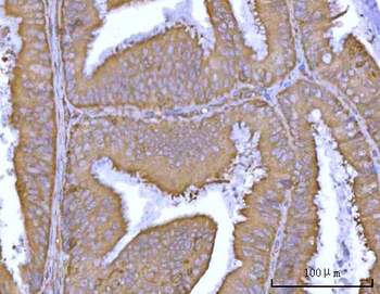
Immunohistochemistry Validation of DR5 in Mouse kidney tissue. Immunohistochemical analysis of paraffin-embedded mouse kidney tissue using anti-DR5 antibody (orb1239348) at 5 µg/ml. Tissue was fixed with formaldehyde and blocked with 10% serum for 1 h at RT; antigen retrieval was by heat mediation with a citrate buffer (pH6). Samples were incubated with primary antibody overnight at 4°C. A goat anti-rabbit IgG H&L (HRP) at 1/250 was used as secondary. Counter stained with Hematoxylin.

KO Validation of DR5 in HCT116 Cells (Han et al., 2015). Anti-cancer drug, Carfilzomib (CFZ), induced up-regulation of DR5 and the expression of DR5 was not detected in DR5-KO HCT 116 cell line with anti-DR5 antibodies (orb1239348).
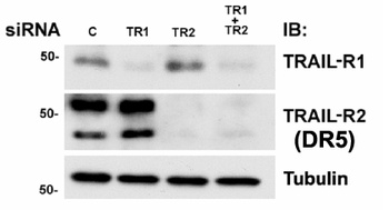
KD Validation of DR5 in MB231 Cells (Rahman et al., 2009). Western blot analysis with anti-DR5 antibodies was performed for DR5 in MB231 cells transfected with control siRNA or DR5 siRNA. DR5 expression was disrupted after DR5 siRNA knockdown.
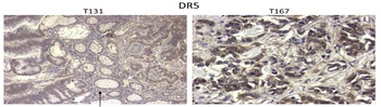
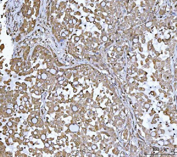
Immunohistochemistry Validation of BIM in Human Colon Tumors (Devetzi et al., 2016). Protein analysis for DR5 by immunohistochemistry with anti-DR5 antibodies in human colon tumors. Strong immunoreactivity is shown for DR5 in T167 patient with colorectal cancer.

Regulated Expression Validation of DR5 in Thyroid Epithelial Cells (Bretz et al., 2002). Immunostaining with anti-DR5 antibodies shows high levels of DR5 expression in untreated cells and cells treated with each of the three cytokines alone or TNFalpha combined with IL-1b. In contrast, treatment with both IFNg and TNFalpha or all three cytokines greatly reduces DR5 staining. The reduction in staining appears most significant in cytoplasmic regions while some staining is maintained in or around the nucleus.
- Item 1 of 8
Anti-DR5/TNFRSF10B Antibody [orb389514]
FC, IHC, WB
Human, Mouse, Rat
Rabbit
Polyclonal
Unconjugated
10 μg, 100 μg - Item 1 of 3
- Item 1 of 3
- Item 1 of 5
Anti-DR5/TNFRSF10B Antibody [orb1728199]
ELISA, FC, IHC, WB
Human, Rat
Rabbit
Polyclonal
Unconjugated
100 μg, 10 μg - Item 1 of 3
Anti-TNFRSF10B antibody(DM115); Rabbit mAb [orb1174053]
ELISA, FC
Human
Rabbit
Monoclonal
Unconjugated
10 μg, 50 μg, 100 μg, 500 μg


![Anti-DR5 [304]](/images//pub/media/catalog/product/NewWebsite/35/orb1463373_1.png)
![Anti-DR5 [304]](/images/pub/media/catalog/product/NewWebsite/35/orb1463373_2.png)
![Anti-DR5 [304]](/images/pub/media/catalog/product/NewWebsite/35/orb1463373_3.png)
![Anti-DR5 [304]](/images//pub/media/catalog/product/NewWebsite/35/orb1463374_1.png)
![Anti-DR5 [304]](/images/pub/media/catalog/product/NewWebsite/35/orb1463374_2.png)
![Anti-DR5 [304]](/images/pub/media/catalog/product/NewWebsite/35/orb1463374_3.png)