You have no items in your shopping cart.
Cart summary
Item 1 of 7
Item 1 of 7
DKK1 Antibody
Catalog Number: orb306404

| Catalog Number | orb306404 |
|---|---|
| Category | Antibodies |
| Description | Rabbit polyclonal antibody to DKK1 |
| Species/Host | Rabbit |
| Clonality | Polyclonal |
| Clone Number | RB52126 |
| Tested applications | IF, IHC-P, WB |
| Reactivity | Human, Mouse, Rat |
| Isotype | Rabbit IgG |
| Dilution range | FACS: 1:25, IHC-P: 1:25, WB: 1:1000-1:4000 |
| Form/Appearance | Purified polyclonal antibody supplied in PBS with 0.09% (W/V) sodium azide. This antibody is purified through a protein A column, followed by peptide affinity purification. |
| Conjugation | Unconjugated |
| MW | 28672 |
| Target | DKK1 |
| UniProt ID | O94907 |
| Storage | Maintain refrigerated at 2-8°C for up to 2 weeks. For long term storage store at -20°C in small aliquots to prevent freeze-thaw cycles |
| Alternative names | anti Dickkopf-related protein 1 antibody, anti Dic Read more... |
| Note | For research use only |
| Expiration Date | 12 months from date of receipt. |

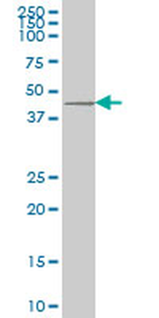
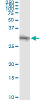
Anti-DKK1 Antibody at 1:4000 dilution + Hela whole cell lysates. Lysates/proteins at 20 µg per lane. Secondary Goat Anti-Rabbit IgG, (H+L), Peroxidase conjugated at 1/10000 dilution. Predicted band size: 29 kDa. Blocking/Dilution buffer: 5% NFDM/TBST.

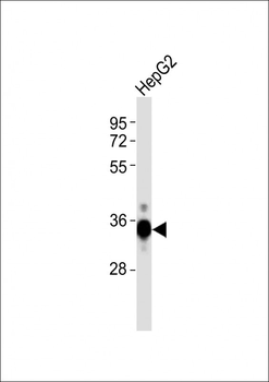
Anti-DKK1 Antibody at 1:1000 dilution + HepG2 whole cell lysates. Lysates/proteins at 20 µg per lane. Secondary Goat Anti-Rabbit IgG, (H+L), Peroxidase conjugated at 1/10000 dilution. Predicted band size: 29 kDa. Blocking/Dilution buffer: 5% NFDM/TBST.

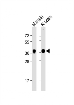
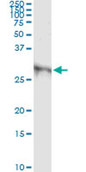
All lanes: Anti-DKK1 Antibody at 1:2000 dilution. Lane 1: Mouse brain lysate. Lane 2: Rat brain lysate. Lysates/proteins at 20 µg per lane. Secondary Goat Anti-Rabbit IgG, (H+L), Peroxidase conjugated at 1/10000 dilution. Predicted band size: 29 kDa. Blocking/Dilution buffer: 5% NFDM/TBST.

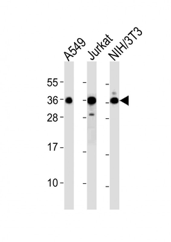
All lanes: Anti-DKK1 Antibody at 1:2000 dilution. Lane 1: A549 whole cell lysates. Lane 2: Jurkat whole cell lysates. Lane 3: NIH/3T3 whole cell lysates. Lysates/proteins at 20 µg per lane. Secondary Goat Anti-Rabbit IgG, (H+L), Peroxidase conjugated at 1/10000 dilution. Predicted band size: 29 kDa. Blocking/Dilution buffer: 5% NFDM/TBST.
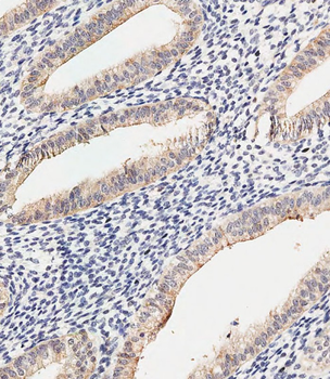
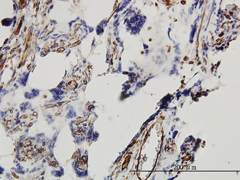
Immunohistochemical analysis of paraffin-embedded Human uterus tissue was performed on the Leica BOND RXm. Tissue was fixed with formaldehyde at room temperature, antigen retrieval was by heat mediation with a EDTA buffer (pH9.0). Samples were incubated with primary antibody (1:500) for 1 hours at room temperature. A undiluted biotinylated CRF Anti-Polyvalent HRP Polymer antibody was used as the secondary antibody.
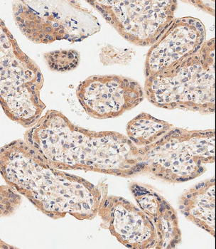
Immunohistochemical analysis of paraffin-embedded Human placenta tissue was performed on the Leica BOND RXm. Tissue was fixed with formaldehyde at room temperature, antigen retrieval was by heat mediation with a EDTA buffer (pH9.0). Samples were incubated with primary antibody (1:500) for 1 hours at room temperature. A undiluted biotinylated CRF Anti-Polyvalent HRP Polymer antibody was used as the secondary antibody.
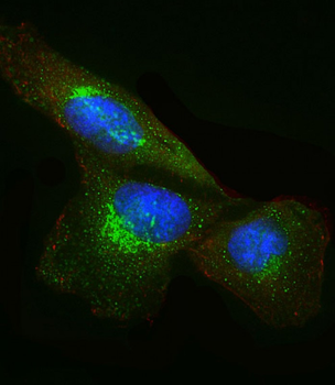
Immunofluorescent analysis of 4% paraformaldehyde-fixed, 0.1% Triton X-100 permeabilized U-251 MG cells labeling DKK1 at 1/25 dilution, followed by Dylight 488-conjugated goat anti-Rabbit IgG secondary antibody at 1/200 dilution (green). Immunofluorescence image showing Cytoplasm and Weak Nucleus staining on U-251 MG cell line. Cytoplasmic actin is detected with Dylight 554 Phalloidin (red). The nuclear counter stain is DAPI (blue).
- Item 1 of 5
Goat anti-DKK1 Antibody [orb18888]
ELISA, IHC, WB
Bovine, Human, Mouse
Goat
Polyclonal
Unconjugated
100 μg - Item 1 of 7
- Item 1 of 4
DKK1 monoclonal antibody (M11), clone 2A5 [orb2291377]
ELISA, IHC-P, IP, WB
Human
Mouse
Monoclonal
Unconjugated
100 μg - Item 1 of 3
- Item 1 of 3
DKK1 monoclonal antibody (M08), clone 2B12 [orb2291378]
ELISA, IHC-P, IP, WB
Human
Mouse
Monoclonal
Unconjugated
100 μg