You have no items in your shopping cart.
Cart summary
Item 1 of 2
Item 1 of 2
Dicer Antibody: FITC
Catalog Number: orb150100

| Catalog Number | orb150100 |
|---|---|
| Category | Antibodies |
| Description | Mouse monoclonal to Dicer (FITC). Dicer is a member of the RNase III family that specifically cleaves double-stranded RNAs to generate microRNAs (miRNAs). After long primary transcript pri-miRNAs are processed to stem-looped pre-miRNAs by Drosha, pre-miRNAs are transported to the cytoplasm and further processed by Dicer to produce 22-nucleotide mature miRNAs. The mature miRNA then becomes a part of the RNA-Induced Silencing Complex (RISC) and can bind to the 3' UTR of the target mRNA.. |
| Species/Host | Mouse |
| Clonality | Monoclonal |
| Clone Number | N167/7 (Formerly sold as S167-7) |
| Tested applications | ICC, IF, IHC |
| Reactivity | Human, Mouse, Rat |
| Isotype | IgG1 |
| Immunogen | Fusion protein amino acids 1638-1899 of mouse Endoribonuclease Dicer |
| Concentration | 1 mg/ml |
| Dilution range | WB (1:1000), ICC/IF (1:100) |
| Conjugation | FITC |
| MW | 215kDa |
| Target | Dicer |
| Entrez | 192119 |
| UniProt ID | Q8R418 |
| NCBI | NP_683750.2 |
| Storage | Conjugated antibodies should be stored according to the product label |
| Buffer/Preservatives | 640.91mM DMSO, 136.36mM Ethanolamine, 9.09mM Sodium Bicarbonate in 90.9% PBS |
| Alternative names | Dicer1 antibody, DCR antibody, DCR1 antibody, DCR- Read more... |
| Note | For research use only |
| Application notes | 1 µg/ml of SMC-416 was sufficient for detection of Dicer in 20 µg of rat brain lysate by colorimetric immunoblot analysis using Goat anti-mouse IgG:HRP as the secondary antibody. |
| Expiration Date | 12 months from date of receipt. |

Immunocytochemistry/Immunofluorescence analysis using Mouse Anti-Dicer Monoclonal Antibody, Clone N167/7. Tissue: Neuroblastoma cells (SH-SY5Y). Species: Human. Fixation: 4% PFA for 15 min. Primary Antibody: Mouse Anti-Dicer Monoclonal Antibody at 1:50 for overnight at 4°C with slow rocking. Secondary Antibody: AlexaFluor 488 at 1:1000 for 1 hour at RT. Counterstain: Phalloidin-iFluor 647 (red) F-Actin stain; Hoechst (blue) nuclear stain at 1:800, 1.6mM for 20 min at RT. (A) Hoechst (blue) nuclear stain. (B) Phalloidin-iFluor 647 (red) F-Actin stain. (C) Dicer Antibody (D) Composite.
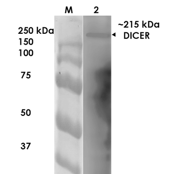
Western Blot analysis of Rat Brain Membrane showing detection of ~215 kDa Dicer protein using Mouse Anti-Dicer Monoclonal Antibody, Clone N167/7. Lane 1: MW Ladder. Lane 2: Rat Brain Membrane. Load: 10 μg. Block: 5% milk. Primary Antibody: Mouse Anti-Dicer Monoclonal Antibody at 1:1000 for 1 hour at RT. Secondary Antibody: Goat Anti-Mouse IgG: HRP at 1:200 for 1 hour at RT. Color Development: TMB solution for 10 min at RT. Predicted/Observed Size: ~215 kDa.

TRBP Rabbit Polyclonal Antibody (FITC) [orb190633]
IF
Bovine, Equine, Human, Porcine, Rat, Sheep
Mouse
Rabbit
Polyclonal
FITC
100 μlDICER1 Rabbit Polyclonal Antibody (FITC) [orb102460]
ICC
Bovine, Equine, Mouse, Porcine, Rat
Human
Rabbit
Polyclonal
FITC
100 μlExportin 5 Rabbit Polyclonal Antibody (FITC) [orb466198]
IF
Bovine, Canine, Equine, Gallus, Human, Porcine, Rabbit, Rat, Sheep
Mouse
Rabbit
Polyclonal
FITC
100 μlDGCR8 Rabbit Polyclonal Antibody (FITC) [orb187392]
ICC, IF
Human, Mouse, Primate, Rat
Rabbit
Polyclonal
FITC
100 μl
