You have no items in your shopping cart.
Cart summary
Item 1 of 4
Item 1 of 4
Cytokeratin 3 antibody
Catalog Number: orb344418

| Catalog Number | orb344418 |
|---|---|
| Category | Antibodies |
| Description | Cytokeratin 3 antibody |
| Species/Host | Mouse |
| Clonality | Monoclonal |
| Clone Number | C11 |
| Tested applications | ELISA, IF, IHC, IP, Multiplex Assay, WB |
| Reactivity | Human |
| Isotype | IgG1 |
| Immunogen | This protein A purified monoclonal antibody was produced by repeated immunizations with purified human cytoskeletal preparations from A431 cells. |
| Concentration | 1.3 mg/mL |
| Dilution range | ELISA: 1:5,000 - 1:20,000, IHC: 1:50 - 1:200, IF: 1:50 - 1:200, IP: 1:100, WB: 1:50 - 1:200 |
| Form/Appearance | Liquid (sterile filtered) |
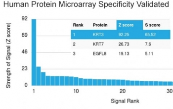
| Purity | This protein A purified mouse monoclonal antibody reacts specifically with keratins from human tissues and derived cell lines. This antibody reacts with keratin (56 kDa), keratin 17 (46 kDa), keratin 18 (45 kDa) and keratin 19 (40 kDa) derived from humans. Cross reactivity with keratins from other sources has not been determined. No reaction is expected against other filament proteins including vimentin, desmin and neurofilament protein. |
| Conjugation | Unconjugated |
| UniProt ID | P48668 |
| NCBI | P48668.3 |
| Storage | Store vial at -20° C prior to opening. Aliquot contents and freeze at -20° C or below for extended storage. Avoid cycles of freezing and thawing. Centrifuge product if not completely clear after standing at room temperature. This product is stable for several weeks at 4° C as an undiluted liquid. Dilute only prior to immediate use. |
| Buffer/Preservatives | 0.01% (w/v) Sodium Azide |
| Alternative names | mouse anti-keratin antibody, mouse anti-cytokerati Read more... |
| Note | For research use only |
| Application notes | Anti-Keratin Antibody has been tested in ELISA, immunohistochemistry, immunofluorescence, immunoblotting and immunoprecipitation. For a positive control use skin, colon carcinoma and squamous granulocyte carcinoma cells. |
| Expiration Date | 12 months from date of receipt. |
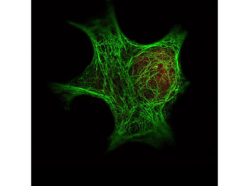
Immunofluorescence Microscopy of Biorbyt's Anti-Keratin antibody (p/n orb344418) was used with Biorbyt's DyLight™ 488 goat anti-mouse [shown in green] to detect Keratin by Immunofluorescence. In the same experiment, Biorbyt's polyclonal Anti-HDAC-1 antibody (p/n orb345508) was used with Atto425 Anti-Rabbit IgG (p/n orb347684) [shown in red] to detect HDAC-1.
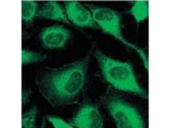
Immunofluorescence using Biorbyt's Mouse Anti-Keratin antibody. Confocal slices of HeLa cells are between 0.5 and 0.6 µM where the image is taken near the bottom of the cell. Use FITC conjugated Goat-a-Mouse IgG [H&L] (p/n orb347377) at 1:2000 dilution for detection.
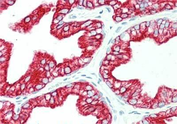
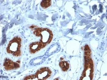
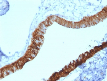
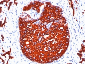
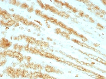
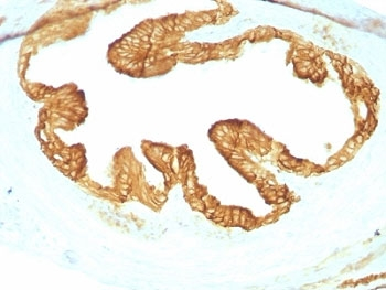
Immunohistochemistry of Mouse anti-Keratin antibody. Tissue: human prostate. Fixation: formalin fixed paraffin embedded. Antigen retrieval: not required. Primary antibody: anti-Keratin antibody at 10 µg/mL for 1 h at RT. Secondary antibody: Peroxidase mouse secondary antibody at 1:10000 for 45 min at RT. Staining: Keratin as precipitated red signal with hematoxylin purple nuclear counterstain.
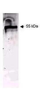
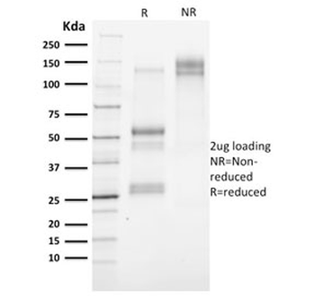
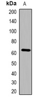
Western blot using Biorbyt's Mouse Anti-Keratin antibody. This antibody recognizes a single 56 kDa band corresponding to human keratin as confirmed by the position of molecular weight markers (not shown). Approximately 100 ng of keratin from human epidermis was applied under reducing conditions to a pre-cast 4-20% iGel from Gradipore Inc. A 1:400 dilution of Mab anti-Keratin was used for 2h followed by detection using a 1:5000 dilution of IRDye™800 conjugated Goat-a-Mouse IgG [H&L].
- Item 1 of 4
- Item 1 of 3
Pan Cytokeratin Antibody Cocktail (Acidic + Basic) [orb749935]
IHC-P
Human, Rat
Mouse
Monoclonal
Unconjugated
20 μg, 100 μg - Item 1 of 3
Cytokeratin 3 antibody [orb182908]
IHC-P
Mouse
Human, Rat
Rabbit
Polyclonal
Unconjugated
50 μl, 100 μl, 200 μl - Item 1 of 1
- Item 1 of 1


Submit a review
Filter by Rating

- 5 stars

- 4 stars
- 3 stars
- 2 stars
- 1 stars
