You have no items in your shopping cart.
Cart summary
Item 1 of 6
Item 1 of 6
CYPA Rabbit Polyclonal Antibody
Catalog Number: orb100498

| Catalog Number | orb100498 |
|---|---|
| Category | Antibodies |
| Description | CYPA Rabbit Polyclonal Antibody |
| Species/Host | Rabbit |
| Clonality | Polyclonal |
| Tested applications | FC, IF, IHC-Fr, IHC-P, WB |
| Predicted Reactivity | Bovine, Canine, Porcine, Rabbit |
| Reactivity | Human, Mouse, Rat |
| Isotype | IgG |
| Immunogen | KLH conjugated synthetic peptide derived from human Cyclophilin A. (101-165/165aa) |
| Antibody Type | Primary Antibody |
| Concentration | 1mg/ml |
| Dilution range | WB=1:500-2000, IHC-P=1:100-500, IHC-F=1:100-500, IF=1:100-500, Flow-Cyt=1μg/Test |
| Form/Appearance | Liquid |
| Conjugation | Unconjugated |
| MW | 18 kDa |
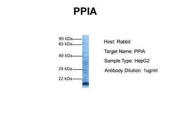
| Target | PPIA |
| UniProt ID | P62937 |
| Storage | Maintain refrigerated at 2-8°C for up to 2 weeks. For long term storage store at -20°C in small aliquots to prevent freeze-thaw cycles. |
| Buffer/Preservatives | 0.01M TBS (pH7.4) with 1% rAlbumin, 0.02% Proclin300 and 50% Glycerol. |
| Alternative names | Cyclophilin A; CyclophilinA; Cyclosporin A binding Read more... |
| Note | For research use only |
| Expiration Date | 12 months from date of receipt. |

Blank control (blue): Hela (fixed with 2% paraformaldehyde (10 min), then permeabilized with 90% ice-cold methanol for 30 min on ice). Primary Antibody: Rabbit Anti-CYPA antibody (orb100498), Dilution: 1 µg in 100 µL 1X PBS containing 0.5% BSA, Isotype Control Antibody: Rabbit IgG (orange), used under the same conditions), Secondary Antibody: Goat anti-rabbit IgG-PE (white blue), Dilution: 1:200 in 1 X PBS containing 0.5% BSA.
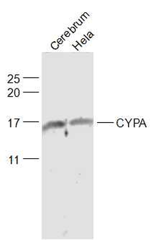
Sample: Cerebrum (Rat) Lysate at 40 ug, Hela (Human) Cell Lysate at 30 ug, Primary: Anti-CYPA (orb100498) at 1/1000 dilution, Secondary: IRDye800CW Goat Anti-Rabbit IgG at 1/20000 dilution, Predicted band size: 18 kD, Observed band size: 17/18 kD.
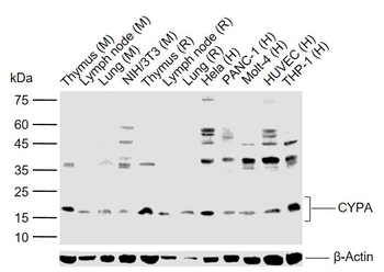
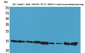
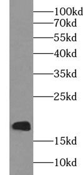
Sample: Lane 1: Mouse Thymus tissue lysates, Lane 2: Mouse Lymph node tissue lysates, Lane 3: Mouse Lung tissue lysates, Lane 4: Mouse NIH/3T3 cell lysates, Lane 5: Rat Thymus tissue lysates, Lane 6: Rat Lymph node tissue lysates, Lane 7: Rat Lung tissue lysates, Lane 8: Human Hela cell lysates, Lane 9: Human PANC-1 cell lysates, Lane 10: Human Molt-4 cell lysates, Lane 11: Human HUVEC cell lysates, Lane 12: Human THP-1 cell lysates, Primary: Anti-CYPA (orb100498) at 1/1000 dilution, Secondary: IRDye800CW Goat Anti-Rabbit IgG at 1/20000 dilution, Predicted band size: 18 kDa, Observed band size: 18 kDa.
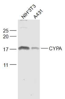
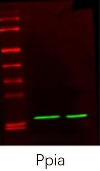
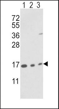
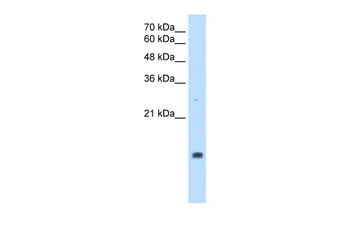
Sample: NIH/3T3 (Mouse) Cell Lysate at 30 ug, A431 (Human) Cell Lysate at 30 ug, Primary: Anti-CYPA (orb100498) at 1/1000 dilution, Secondary: IRDye800CW Goat Anti-Rabbit IgG at 1/20000 dilution, Predicted band size: 18 kD, Observed band size: 18 kD.
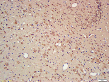
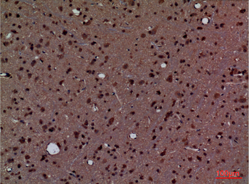
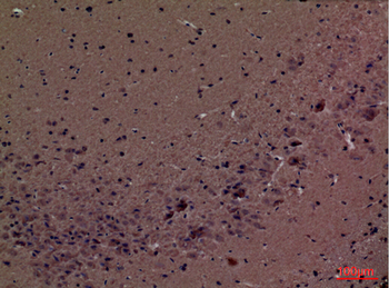
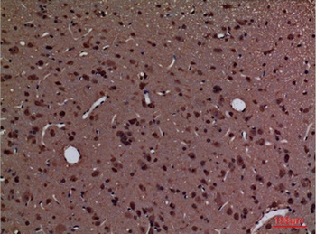
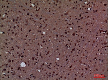
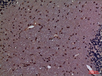
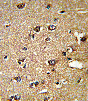
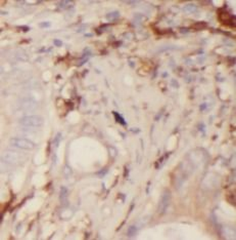
Tissue/cell: rat brain tissue, 4% Paraformaldehyde-fixed and paraffin-embedded, Antigen retrieval: citrate buffer (0.01M, pH 6.0), Boiling bathing for 15 min, Block endogenous peroxidase by 3% Hydrogen peroxide for 30 min, Blocking buffer (normal goat serum) at 37℃ for 20 min, Incubation: Anti-CYPA Polyclonal Antibody, Unconjugated (orb100498) 1:200, overnight at 4°C, followed by conjugation to the secondary antibody and DAB staining.
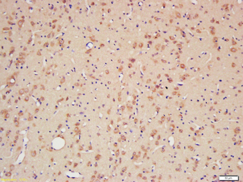
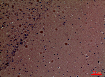
Tissue/cell: rat brain tissue, 4% Paraformaldehyde-fixed and paraffin-embedded, Antigen retrieval: citrate buffer (0.01M, pH 6.0), Boiling bathing for 15 min, Block endogenous peroxidase by 3% Hydrogen peroxide for 30 min, Blocking buffer (normal goat serum) at 37℃ for 20 min, Incubation: Anti-CYPA Polyclonal Antibody, Unconjugated (orb100498) 1:200, overnight at 4°C, followed by conjugation to the secondary antibody and DAB staining.
- Item 1 of 7
- Item 1 of 4
- Item 1 of 4
PPIA Antibody (N-term) [orb1931319]
FC, IHC-P, WB
Hamster, Porcine, Rabbit, Rat
Human, Mouse
Rabbit
Polyclonal
Unconjugated
100 μl, 50 μl - Item 1 of 3
PPIA Rabbit Polyclonal Antibody [orb581362]
IHC, WB
Bovine, Canine, Equine, Goat, Guinea pig, Mouse, Rabbit, Rat, Sheep
Human
Rabbit
Polyclonal
Unconjugated
100 μl - Item 1 of 3
PPIA Antibody [orb626170]
ELISA, FC, IF, IHC, WB
Human, Mouse, Rat
Rabbit
Polyclonal
Unconjugated
100 μg, 50 μg