You have no items in your shopping cart.
Cart summary
Item 1 of 3
Item 1 of 3
CYP2B6 Antibody (Center)
Catalog Number: orb1439557

| Catalog Number | orb1439557 |
|---|---|
| Category | Antibodies |
| Description | Rabbit polyclonal antibody to CYP2B6. |
| Species/Host | Rabbit |
| Clonality | Polyclonal |
| Clone Number | RB16992 |
| Tested applications | IF, IHC-P, WB |
| Reactivity | Human |
| Isotype | Rabbit IgG |
| Immunogen | Synthetic Peptide |
| Dilution range | IF: 1:25, WB: 1:2000, IHC-P: 1:10~50 |
| Form/Appearance | Purified polyclonal antibody supplied in PBS with 0.09% (W/V) sodium azide. This antibody is purified through a protein A column, followed by peptide affinity purification. |
| Conjugation | Unconjugated |
| MW | 56278 |
| Target | CYP2B6 |
| UniProt ID | P20813 |
| NCBI | NP_000758.1 |
| Storage | Maintain refrigerated at 2-8°C for up to 2 weeks. For long term storage store at -20°C in small aliquots to prevent freeze-thaw cycles |
| Alternative names | Cytochrome P450 2B6, 11413-,4-cineole 2-exo-monoox Read more... |
| Note | For research use only |
| Expiration Date | 12 months from date of receipt. |

All lanes: Anti-CYP2B6 Antibody (Center) at 1:2000 dilution. Lane 1: 293 whole cell lysate. Lane 2: Jurkat whole cell lysate.Lysates/proteins at 20 µg per lane. Secondary Goat Anti-Rabbit IgG, (H+L), Peroxidase conjugated at 1/10000 dilution. Predicted band size: 56 kDa. Blocking/Dilution buffer: 5% NFDM/TBST.

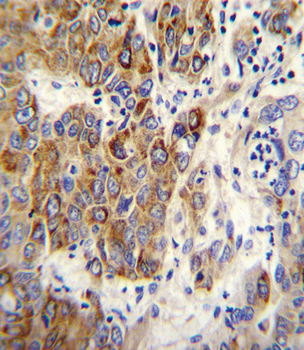
CYP2B6 Antibody (Center) immunohistochemistry analysis in formalin fixed and paraffin embedded human hepatocarcinoma followed by peroxidase conjugation of the secondary antibody and DAB staining. This data demonstrates the use of CYP2B6 Antibody (Center) for immunohistochemistry. Clinical relevance has not been evaluated.
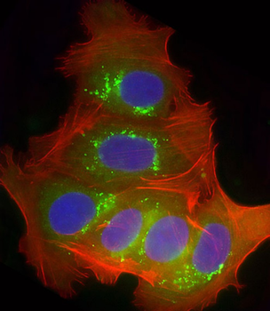
Immunofluorescent analysis of 4% paraformaldehyde-fixed, 0.1% Triton X-100 permeabilized MCF-7 (human breast cancer cell line) cells labeling CYP2B6 at 1/25 dilution, followed by Dylight 488-conjugated goat anti-rabbit IgG secondary antibody at 1/200 dilution (green). Immunofluorescence image showing endosomes staining on MCF-7 cell line. Cytoplasmic actin is detected with Dylight 554 Phalloidin at 1/100 dilution (red). The nuclear counter stain is DAPI (blue).
- Item 1 of 3