You have no items in your shopping cart.
Cart summary
Item 1 of 6
Item 1 of 6
CTSA Antibody (N-term)
Catalog Number: orb1938960

| Catalog Number | orb1938960 |
|---|---|
| Category | Antibodies |
| Description | Affinity Purified Rabbit Polyclonal Antibody (Pab) |
| Species/Host | Rabbit |
| Clonality | Polyclonal |
| Clone Number | RB23380 |
| Tested applications | IHC-P, WB |
| Reactivity | Human, Mouse |
| Isotype | Rabbit IgG |
| Dilution range | WB: 1:1000, WB: 1:2000, WB: 1:1000, WB: 1:1000, IHC-P-Leica: 1:500, IHC-P-Leica: 1:500 |
| Form/Appearance | Purified polyclonal antibody supplied in PBS with 0.09% (W/V) sodium azide. This antibody is purified through a protein A column, followed by peptide affinity purification. |
| Conjugation | Unconjugated |
| MW | 54466 Da |
| Target | This CTSA antibody is generated from rabbits immunized with a KLH conjugated synthetic peptide between 18-45 amino acids from the N-terminal region of human CTSA. |
| UniProt ID | P10619 |
| NCBI | NP_000299.2, NP_001121167.1, NP_001161066.1 |
| Storage | Maintain refrigerated at 2-8°C for up to 2 weeks. For long term storage store at -20°C in small aliquots to prevent freeze-thaw cycles |
| Alternative names | Lysosomal protective protein, Carboxypeptidase C, Read more... |
| Note | For research use only |
| Expiration Date | 12 months from date of receipt. |

Anti-CTSA Antibody (N-term) at 1:2000 dilution + 293 whole cell lysate.Lysates/proteins at 20 µg per lane. Secondary Goat Anti-Rabbit IgG, (H+L), Peroxidase conjugated at 1/10000 dilution. Predicted band size: 54 kDa. Blocking/Dilution buffer: 5% NFDM/TBST.

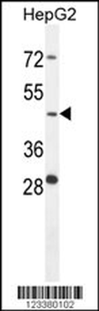
CTSA Antibody (N-term) western blot analysis in HepG2 cell line lysates (35 ug/lane). This demonstrates the CTSA antibody detected the CTSA protein (arrow).
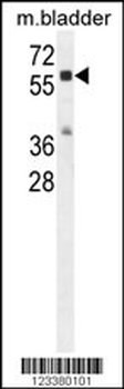
CTSA Antibody (N-term) western blot analysis in mouse bladder tissue lysates (35 ug/lane). This demonstrates the CTSA antibody detected the CTSA protein (arrow).

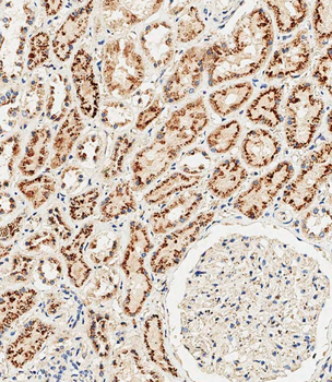
Immunohistochemical analysis of paraffin-embedded Human kidney tissue performed on the Leica BOND RXm. Tissue was fixed with formaldehyde at room temperature, antigen retrieval was by heat mediation with a EDTA buffer (pH9.0). Samples were incubated with primary antibody (1:500) for 1 hours at room temperature. A undiluted biotinylated CRF Anti-Polyvalent HRP Polymer antibody was used as the secondary Antibody.
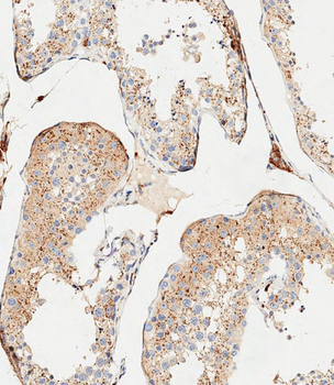
Immunohistochemical analysis of paraffin-embedded Human testis tissue performed on the Leica BOND RXm. Tissue was fixed with formaldehyde at room temperature, antigen retrieval was by heat mediation with a EDTA buffer (pH9.0). Samples were incubated with primary antibody (1:500) for 1 hours at room temperature. A undiluted biotinylated CRF Anti-Polyvalent HRP Polymer antibody was used as the secondary Antibody.
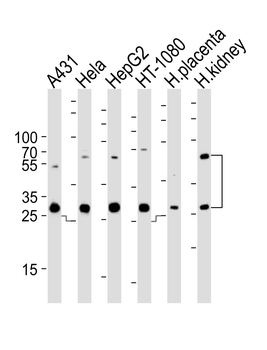
Western blot analysis of lysates from A431, Hela, HepG2, HT-1080 cell line and human placenta, kidney tissue lysate (from left to right), using CTSA Antibody (N-term). diluted at 1:1000 at each lane. A goat anti-rabbit IgG H&L (HRP) at 1:10000 dilution was used as the secondary Antibody. Lysates at 35 ug per lane.
- Item 1 of 6