You have no items in your shopping cart.
Cart summary
Item 1 of 4
Item 1 of 4
CTBP1 Antibody (C-term)
Catalog Number: orb1788395

| Catalog Number | orb1788395 |
|---|---|
| Category | Antibodies |
| Description | Affinity Purified Rabbit Polyclonal Antibody (Pab) |
| Species/Host | Rabbit |
| Clonality | Polyclonal |
| Tested applications | IF, WB |
| Reactivity | Human, Mouse, Rat |
| Isotype | Rabbit IgG |
| Immunogen | 413-440 aa |
| Dilution range | IF: 1:10~50, IF: 1:10~50, WB: 1:1000, WB: 1:2000 |
| Form/Appearance | Purified polyclonal antibody supplied in PBS with 0.09% (W/V) sodium azide. This antibody is purified through a protein A column, followed by peptide affinity purification. |
| Conjugation | Unconjugated |
| MW | 47535 Da |
| Target | This CTBP1 antibody is generated from rabbits immunized with a KLH conjugated synthetic peptide between 413-440 amino acids from the C-terminal region of human CTBP1. |
| UniProt ID | Q13363 |
| NCBI | NP_001319.1, NP_001012632.1 |
| Storage | Maintain refrigerated at 2-8°C for up to 2 weeks. For long term storage store at -20°C in small aliquots to prevent freeze-thaw cycles |
| Alternative names | CTBP1; CTBP; C-terminal-binding protein 1 Read more... |
| Note | For research use only |
| Expiration Date | 12 months from date of receipt. |

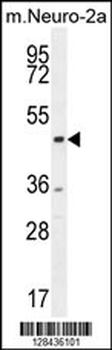
Western blot analysis of lysates from Jurkat cell line, mouse brain, rat brain tissue lysate, Raji, Hela cell line (from left to right), using CTBP1 Antibody (C-term). Diluted at 1:1000 at each lane. A goat anti-rabbit IgG H&L (HRP) at 1:10000 dilution was used as the secondary antibody. Lysates at 20ug per lane.

All lanes: Anti-CTBP1 Antibody (C-term) at 1:2000 dilution. Lane 1: HepG2 whole cell lysate. Lane 2: Jurkat whole cell lysate. Lane 3: Raji whole cell lysate. Lane 4: Hela whole cell lysate. Lane 5: mouse brain lysate. Lysates/proteins at 20 µg per lane. Secondary Goat Anti-Rabbit IgG, (H+L), Peroxidase conjugated at 1/10000 dilution. Predicted band size: 48 kDa. Blocking/Dilution buffer: 5% NFDM/TBST.
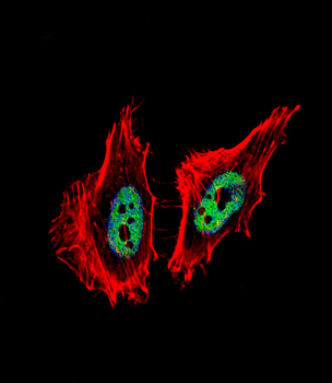
Fluorescent confocal image of Hela cell stained with CTBP1 Antibody (C-term). Hela cells were fixed with 4% PFA (20 min), permeabilized with Triton X-100 (0.1%, 10 min), then incubated with CTBP1 primary antibody (1:25, 1 h at 37°C). For secondary antibody, Alexa Fluor 488 conjugated donkey anti-rabbit antibody (green) was used (1:400, 50 min at 37°C).Cytoplasmic actin was counterstained with Alexa Fluor 555 (red) conjugated Phalloidin (7units/ml, 1 h at 37°C). Nuclei were counterstained with DAPI (blue) (10 µg/ml, 10 min).CTBP1 immunoreactivity is localized to nucleus significantly.
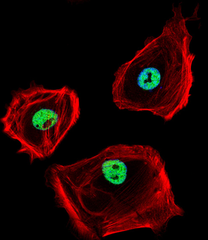
Fluorescent confocal image of SK-BR-3 cell stained with CTBP1 Antibody (C-term). SK-BR-3 cells were fixed with 4% PFA (20 min), permeabilized with Triton X-100 (0.1%, 10 min), then incubated with CTBP1 primary antibody (1:25, 1 h at 37°C). For secondary antibody, Alexa Fluor 488 conjugated donkey anti-rabbit antibody (green) was used (1:400, 50 min at 37°C).Cytoplasmic actin was counterstained with Alexa Fluor 555 (red) conjugated Phalloidin (7units/ml, 1 h at 37°C). Nuclei were counterstained with DAPI (blue) (10 µg/ml, 10 min).CTBP1 immunoreactivity is localized to nucleus significantly.
- Item 1 of 2
- Item 1 of 1