You have no items in your shopping cart.
Cart summary
Item 1 of 5
Item 1 of 5
CSF1R Antibody
Catalog Number: orb1926522

| Catalog Number | orb1926522 |
|---|---|
| Category | Antibodies |
| Description | Purified Mouse Monoclonal Antibody (Mab) |
| Species/Host | Mouse |
| Clonality | Monoclonal |
| Clone Number | 1486CT328.53.37 |
| Tested applications | FC, IHC-P, WB |
| Reactivity | Human |
| Isotype | IgG1,k |
| Dilution range | WB: 1:2000, WB: 1:4000, IHC-P: 1:25, IHC-P: 1:25, FC: 1:25 |
| Form/Appearance | Purified monoclonal antibody supplied in PBS with 0.09% (W/V) sodium azide. This antibody is purified through a protein G column, followed by dialysis against PBS. |
| Conjugation | Unconjugated |
| MW | 107984 Da |
| Target | This CSF1R antibody is generated from a mouse immunized with a recombinant protein. |
| UniProt ID | P07333 |
| Storage | Maintain refrigerated at 2-8°C for up to 2 weeks. For long term storage store at -20°C in small aliquots to prevent freeze-thaw cycles |
| Alternative names | Macrophage colony-stimulating factor 1 receptor, C Read more... |
| Note | For research use only |
| Expiration Date | 12 months from date of receipt. |

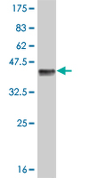
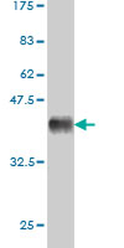
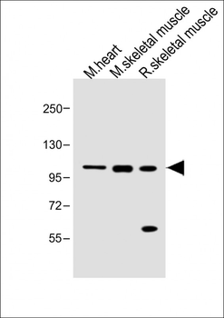
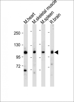
Anti-CSF1R Antibodyat 1:2000 dilution + human placenta lysates. Lysates/proteins at 20 μg per lane. Secondary Goat Anti-mouse IgG, (H+L), Peroxidase conjugated at 1/10000 dilution. Predicted band size: 108 kDa. Blocking/Dilution buffer: 5% NFDM/TBST.
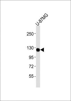
Anti-CSF1R Antibodyat 1:4000 dilution + U-87MG whole cell lysates. Lysates/proteins at 20 μg per lane. Secondary Goat Anti-mouse IgG, (H+L), Peroxidase conjugated at 1/10000 dilution. Predicted band size: 108 kDa. Blocking/Dilution buffer: 5% NFDM/TBST.
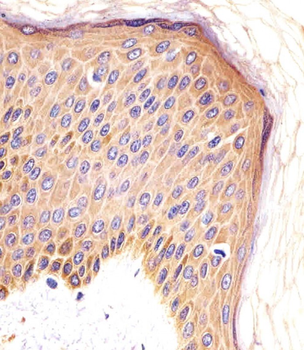
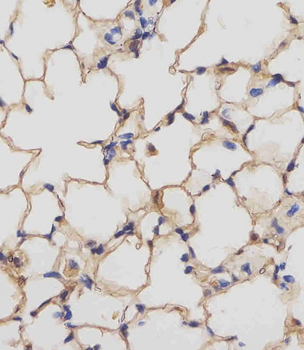
Staining CSF1R in human skin sections by Immunohistochemistry (IHC-P - paraformaldehyde-fixed, paraffin-embedded sections). Tissue was fixed with formaldehyde and blocked with 3% BSA for 0.5 hour at room temperature; antigen retrieval was by heat mediation with a citrate buffer (pH6). Samples were incubated with primary antibody (1/25) for 1 hours at 37°C. A undiluted biotinylated goat polyvalent antibody was used as the secondary antibody.
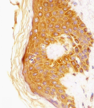
Staining CSF1R in human skin sections by Immunohistochemistry (IHC-P - paraformaldehyde-fixed, paraffin-embedded sections). Tissue was fixed with formaldehyde and blocked with 3% BSA for 0.5 hour at room temperature; antigen retrieval was by heat mediation with a citrate buffer (pH6). Samples were incubated with primary antibody (1/25) for 1 hours at 37°C. A undiluted biotinylated goat polyvalent antibody was used as the secondary antibody.
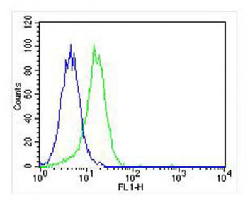
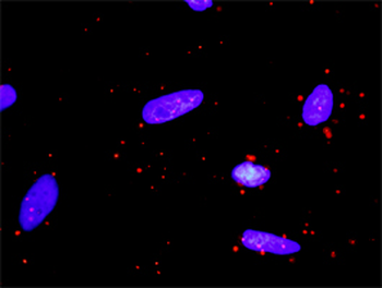
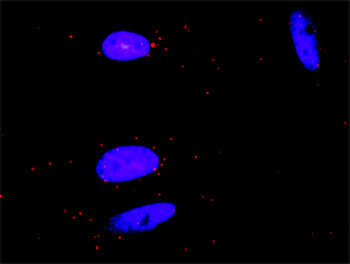
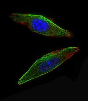
Overlay histogram showing U-87MG cells stained (green line). The cells were fixed with 2% paraformaldehyde (10 min). The cells were then icubated in 2% bovine serum albumin to block non-specific protein-protein interactions followed by the antibody (1:25 dilution) for 60 min at 37°C. The secondary antibody used was Goat-Anti-mouse IgG, DyLight 488 Conjugated Highly Cross-Adsorbed at 1/400 dilution for 40 min at 37°C. Isotype control antibody (blue line) was rabbit IgG (1 μg/1x10^6 cells) used under the same conditions. Acquisition of > 10000 events was performed.
- Item 1 of 6
- Item 1 of 4
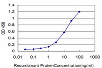
CSF1R monoclonal antibody (M02), clone 3G12 [orb2294065]
ELISA, PLA, WB
Human
Mouse
Monoclonal
Unconjugated
100 μg - Item 1 of 4
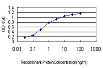
CSF1R monoclonal antibody (M01), clone 1G4 [orb2294066]
ELISA, PLA, WB
Human
Mouse
Monoclonal
Unconjugated
100 μg - Item 1 of 4
CSF1R (Ab-809) Antibody [orb685574]
ELISA, IF, IHC, WB
Human, Mouse
Rabbit
Polyclonal
Unconjugated
100 μl - Item 1 of 5
Mouse Csf1r Antibody (C-term) [orb1936300]
IF, IHC-P, WB
Mouse, Rat
Rabbit
Polyclonal
Unconjugated
100 μl, 50 μl