You have no items in your shopping cart.
Cart summary
Item 1 of 3
Item 1 of 3
CREB (phospho-S133) antibody
Catalog Number: orb345380

| Catalog Number | orb345380 |
|---|---|
| Category | Antibodies |
| Description | CREB (phospho-S133) antibody |
| Species/Host | Rabbit |
| Clonality | Polyclonal |
| Tested applications | ELISA, IHC, WB |
| Reactivity | Human, Mouse, Rat |
| Isotype | IgG |
| Immunogen | CREB phospho peptide corresponding to amino acid residues 122-147 of the human protein conjugated to Keyhole Limpet Hemocyanin (KLH). |
| Concentration | 0.064 mg/mL |
| Dilution range | ELISA: 1:1,000 - 1:6,000, IHC: 20 µg/ml, WB: 1:500 - 1:2,000 |
| Form/Appearance | Liquid (sterile filtered) |
| Purity | Anti-CREB pS133 was affinity purified from monospecific antiserum by immunoaffinity purification. Antiserum was first sequentially pre-adsorbed against a control E.coli lysate, nickel-purified recombinant CREB and the non-phosphorylated form of the immunizing peptide. The resultant depleted antiserum was then purified against the phosphorylated form of the immunizing peptide. This phospho specific polyclonal antibody is specific for phosphorylated pS133 of human CREB. Reactivity with non-phosphorylated CREB is minimal. This antibody does show cross reactivity with pS133 phosphorylated CREB from mouse and rat. |
| Conjugation | Unconjugated |
| UniProt ID | P16220 |
| NCBI | CAG28545.1 |
| Storage | Store vial at -20° C prior to opening. Aliquot contents and freeze at -20° C or below for extended storage. Avoid cycles of freezing and thawing. Centrifuge product if not completely clear after standing at room temperature. This product is stable for several weeks at 4° C as an undiluted liquid. Dilute only prior to immediate use. |
| Buffer/Preservatives | 0.01% (w/v) Sodium Azide |
| Alternative names | rabbit anti-CREB pS133 Antibody, Cyclic AMP-respon Read more... |
| Note | For research use only |
| Application notes | Anti-CREB pS133 antibody reacts with phosphorylated human CREB at pS133 and shows minimal reactivity by western blot with non-phosphorylated CREB and minimal reactivity (1%) by ELISA against the non-phosphorylated form of the immunizing peptide. This antibody was assayed against a variety of tissues including fibroblasts and B-cell lysates. Bands of 46 and 43 kDa corresponding to phosphorylated CREB are observed in western blots. Anti-CREB pS133 is suitable for the detection by immunoblot of phosphorylated human, mouse and rat CREB. No cross-reactivity occurs with non-phosphorylated CREB. For immunohistochemistry, formalin fixed, paraffin embedded human tissue shows moderate to strong nuclear staining in a variety of cells with minimal background staining. Although not tested this antibody is likely functional by FACS and immunoprecipitation. |
| Expiration Date | 12 months from date of receipt. |
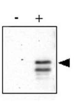
Anti-CREB pS133 was used to detect phosphorylated CREB by western blot at ~46 kDa. Recombinant His-tagged human CREB was produced in E.coli and purified by metal affinity chromatography. An aliquot of purified CREB was phosphorylated in-vitro using Protein Kinase A and ATP. Western blot of control (-) and in-vitro phosphorylated CREB (+) was used to show that the antibody reacts specifically with the phosphorylated form. Pan reactive CREB (Biorbyt # orb750462) reacts equally with both non-phosphorylated and phosphorylated CREB (not shown). Detection occurs using a 1:500 dilution of antibody followed by 1:5000 dilution of HRP Goat-a-Rabbit IgG with visualization via ECL. Film exposure was approximately 1'. Other detection systems will yield similar results.
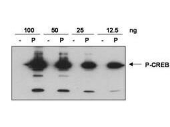
Anti-CREB pS133 was used to detect phosphorylated CREB by western blot. Recombinant His-tagged human CREB was produced in E.coli and purified by metal affinity chromatography. An aliquot of purified CREB was phosphorylated in-vitro using Protein Kinase A and ATP. Western blot of indicated amounts (100 ng, 50 ng, 25 ng, 12.5 ng) of control (-) and in-vitro phosphorylated CREB (P) were loaded to show that the antibody reacts specifically with the phosphorylated form. Blots were blocked in 5% milk in TBS+ 0.1% Tween-20 (TBST-M) overnight at 4°C. Detection occurs using a 1:500 dilution of antibody diluted in TBST-M and incubated at room temperature with rocking for 1 hour. Blots were rinsed 6X with TBST and incubated with goat anti-rabbit-HRP at 1:5000 in TBST-M at room temperature for 45 min. Blots were again rinsed 6X with TBST and then processed using ECL reagent. Exposure time: 1 min.
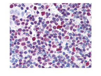
Biorbyt's affinity purified anti-CREB pS133 antibody was used at 20 µg/ml to detect signal in a variety of tissues including multi-human, multi-brain and multi-cancer slides. This image shows moderate to strong nuclear staining of tonsillar lymphocytes. Tissue was formalin-fixed and paraffin embedded. The image shows localization of the antibody as the precipitated red signal, with a hematoxylin purple nuclear counterstain.
- Item 1 of 5
CREB1 (phospho-Ser133) antibody [orb10462]
FC, IHC-P, WB
Bovine, Canine, Gallus, Porcine, Sheep
Human, Mouse, Rat
Rabbit
Polyclonal
Unconjugated
200 μl, 50 μl, 100 μl - Item 1 of 2
CREB (phospho-S133) antibody [orb1474549]
IH, IP, WB
Human, Rat
Rabbit
Monoclonal
Unconjugated
30 μl, 100 μl, 200 μl - Item 1 of 2
CREB1 (Phospho-S133) antibody [orb537099]
ICC, IF, IHC, WB
Human, Mouse, Rat
Polyclonal
Unconjugated
50 μl, 100 μl, 200 μl - Item 1 of 1
CREB1 (Phospho-S133) antibody [orb593296]
IHC, WB
Human, Mouse, Rat
Polyclonal
Unconjugated
200 μl, 100 μl, 50 μl 
CREB1 (Phospho-S133) antibody [orb571529]
ELISA, IHC, IP, WB
Human, Mouse, Rat
Rabbit
Polyclonal
Unconjugated
50 μg, 100 μg


Submit a review
Filter by Rating

- 5 stars

- 4 stars
- 3 stars
- 2 stars
- 1 stars
