You have no items in your shopping cart.
Cart summary
Item 1 of 2
Item 1 of 2
Cpn10 Antibody: PerCP
Catalog Number: orb152104

| Catalog Number | orb152104 |
|---|---|
| Category | Antibodies |
| Description | Rabbit polyclonal to Cpn10 (PerCP). Chaperonin 10, otherwise known as Cpn10, (groES in E. coli) make up a family of small heart shock proteins with an approximate molecular mass of 10kDa (Hsp10s). Cpn10 acts as a co-chaperone and interacts with the Hsp60 family to promote proper folding of polypeptides. Cpn10 and Cpn60 both exhibit sevenfold axis of symmetry and function as a team in the protein folding and assembly process. Cpn10 has been located in human platelets, but is also present in human maternal serum. It has been reported that human Cpn10 is identical with early pregnancy factor, which is involved in control over cell growth and development. This identification suggest that Cpn10 may act like a hormone in stressful situations such as pregnancy.. |
| Species/Host | Rabbit |
| Clonality | Polyclonal |
| Tested applications | ELISA, ICC, IF, IHC, WB |
| Reactivity | Bovine, Canine, Frog, Guinea pig, Human, Mouse, Porcine, Rabbit, Rat, Sheep |
| Immunogen | Human Cpn10 peptide AA 91-101 |
| Concentration | 1 mg/ml |
| Dilution range | WB (1:1000), IHC (1:100) |
| Conjugation | PerCP |
| MW | 10kDa |
| Target | Cpn10 |
| Entrez | 3336 |
| UniProt ID | P61604 |
| NCBI | NP_002148.1 |
| Storage | Conjugated antibodies should be stored according to the product label |
| Buffer/Preservatives | 95.64mM Phosphate, 2.48mM MES and 2mM EDTA |
| Alternative names | 10kDa Chaperonin antibody, Chaperonin 10 antibody, Read more... |
| Note | For research use only |
| Application notes | 1 µg/ml of SPC-193 was sufficient for detection of Cpn10 in 10 µg of Hela lysates by colorimetric immunoblot analysis using Goat anti-rabbit IgG:HRP as the secondary antibody. |
| Expiration Date | 12 months from date of receipt. |

Immunohistochemistry analysis using Rabbit Anti-Cpn10 Polyclonal Antibody. Tissue: backskin. Species: Mouse. Fixation: Bouin's Fixative Solution. Primary Antibody: Rabbit Anti-Cpn10 Polyclonal Antibody at 1:100 for 1 hour at RT. Secondary Antibody: FITC Goat Anti-Rabbit (green) at 1:50 for 1 hour at RT. Localization: Strong basal layer punctate staining in the cytoplasm, weakens as it ascends to upper epidermis.
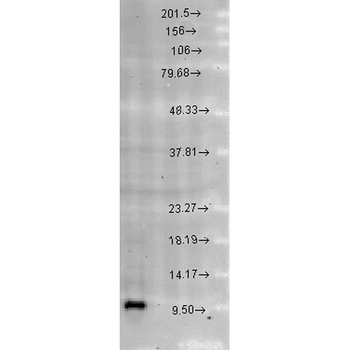
Western blot analysis of Rat brain cell lysates showing detection of Cpn10 protein using Rabbit Anti-Cpn10 Polyclonal Antibody. Load: 15 μgprotein. Block: 1.5% BSA for 30 minutes at RT. Primary Antibody: Rabbit Anti-Cpn10 Polyclonal Antibody at 1:1000 for 2 hours at RT. Secondary Antibody: Donkey Anti-Rabbit IgG: HRP for 1 hour at RT.

CPN10 Rabbit Polyclonal Antibody (PerCP-Cy5.5) [orb2416706]
IF
A. thaliana, Plant
Rabbit
Polyclonal
PerCP/Cy5.5
100 μlCPN10 Rabbit Polyclonal Antibody (PerCP) [orb2416707]
IF
A. thaliana, Plant
Rabbit
Polyclonal
PerCP
100 μlHSPE1 Rabbit Polyclonal Antibody (PerCP-Cy5.5) [orb2395586]
IF
Mouse, Rat
Human
Rabbit
Polyclonal
PerCP/Cy5.5
100 μl
