You have no items in your shopping cart.
Cart summary

| Catalog Number | orb764909 |
|---|---|
| Category | Antibodies |
| Description | Rabbit polyclonal antibody to Cox2 |
| Clonality | Polyclonal |
| Tested applications | ELISA, IF, IHC-P, WB |
| Reactivity | Human, Mouse, Rat |
| Isotype | IgG |
| Immunogen | The antiserum was produced against synthesized peptide derived from human Cox2. AA range:555-604 |
| Concentration | 1 mg/ml |
| Dilution range | Western Blot: 1/500 - 1/2000. Immunohistochemistry: 1/100 - 1/300. Immunofluorescence: 1/200 - 1/1000. ELISA: 1/20000. Not yet tested in other applications. |
| Conjugation | Unconjugated |
| MW | 80 |
| Source | Rabbit |
| Storage | Maintain refrigerated at 2-8°C for up to 2 weeks. For long term storage store at -20°C in small aliquots to prevent freeze-thaw cycles. |
| Buffer/Preservatives | PBS with 0.02% sodium azide and 50% glycerol pH 7.4. |
| Alternative names | Anti-PTGS2 antibody, anti-COX2 antibody, anti-Pros Read more... |
| Note | For research use only |
| Expiration Date | 12 months from date of receipt. |

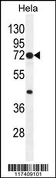
Western blot analysis of lysates from 1) A549, 2) HELA , 3) MCF-7 cells, (Green) primary antibody was diluted at 1:1000, 4°C over night, secondary antibody was diluted at 1:10000, 37°C 1 hour. (Red) Actin β Monoclonal Antibody (5B7) antibody was diluted at 1:5000 as loading control, 4°C over night, secondary antibody was diluted at 1:10000, 37°C 1 hour.

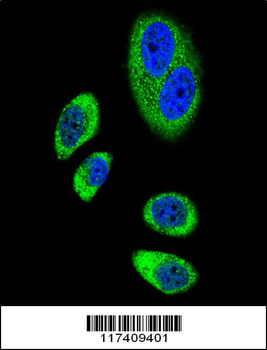
Immunofluorescence analysis of human-liver-cancer tissue. 1, Cox-2 Polyclonal Antibody (red) was diluted at 1:200 (4°C, overnight). 2, Cy3 labled Secondary antibody was diluted at 1:300 (room temperature, 50min). 3, Picture B: DAPI (blue) 10min. Picture A:Target. Picture B: DAPI. Picture C: merge of A + B.

Immunofluorescence analysis of human-liver-cancer tissue. 1, Cox-2 Polyclonal Antibody (red) was diluted at 1:200 (4°C, overnight). 2, Cy3 labled Secondary antibody was diluted at 1:300 (room temperature, 50min). 3, Picture B: DAPI (blue) 10min. Picture A:Target. Picture B: DAPI. Picture C: merge of A + B.

Immunofluorescence analysis of human-lung-cancer tissue. 1, Cox-2 Polyclonal Antibody (red) was diluted at 1:200 (4°C, overnight). 2, Cy3 labled Secondary antibody was diluted at 1:300 (room temperature, 50min). 3, Picture B: DAPI (blue) 10min. Picture A:Target. Picture B: DAPI. Picture C: merge of A + B.
- Item 1 of 4
PTGS2 Antibody (Center P378) [orb1938116]
IF, IHC-P, WB
Human
Rabbit
Polyclonal
Unconjugated
50 μl, 100 μl 
Cyclooxygenase 2 Rabbit pAb [orb1924181]
IHC-P, WB
Human, Mouse, Rat
Polyclonal
Unconjugated
100 μl, 50 μl