You have no items in your shopping cart.
Cart summary
Item 1 of 4
Item 1 of 4
COX-1 Antibody
Catalog Number: orb1247911

| Catalog Number | orb1247911 |
|---|---|
| Category | Antibodies |
| Description | COX-1 Antibody |
| Species/Host | Goat |
| Clonality | Polyclonal |
| Tested applications | ELISA, IF, WB |
| Reactivity | Human, Mouse |
| Immunogen | The immunogen for this antibody is: C-QDDGPAVERPSTEL |
| Antibody Type | Primary Antibody |
| Concentration | 500 ug/mL |
| Form/Appearance | Liquid |
| Conjugation | Unconjugated |
| Target | PTGS1 |
| UniProt ID | P23219 |
| NCBI | NP_542158.1, NP_000953.2 |
| Storage | Maintain refrigerated at 2-8°C for up to 2 weeks. For long term storage store at -20°C in small aliquots to prevent freeze-thaw cycles. |
| Buffer/Preservatives | Supplied at 0.5 mg/ml in Tris saline, 0.02% sodium azide, pH 7.3 with 0.5% bovine serum albumin. Aliquot and store at -20°C. Minimize freezing and thawing. |
| Alternative names | PTGS1, COX1, COX3, PHS1, PCOX1, PGHS1, PTGHS, PGG/ Read more... |
| Note | For research use only |
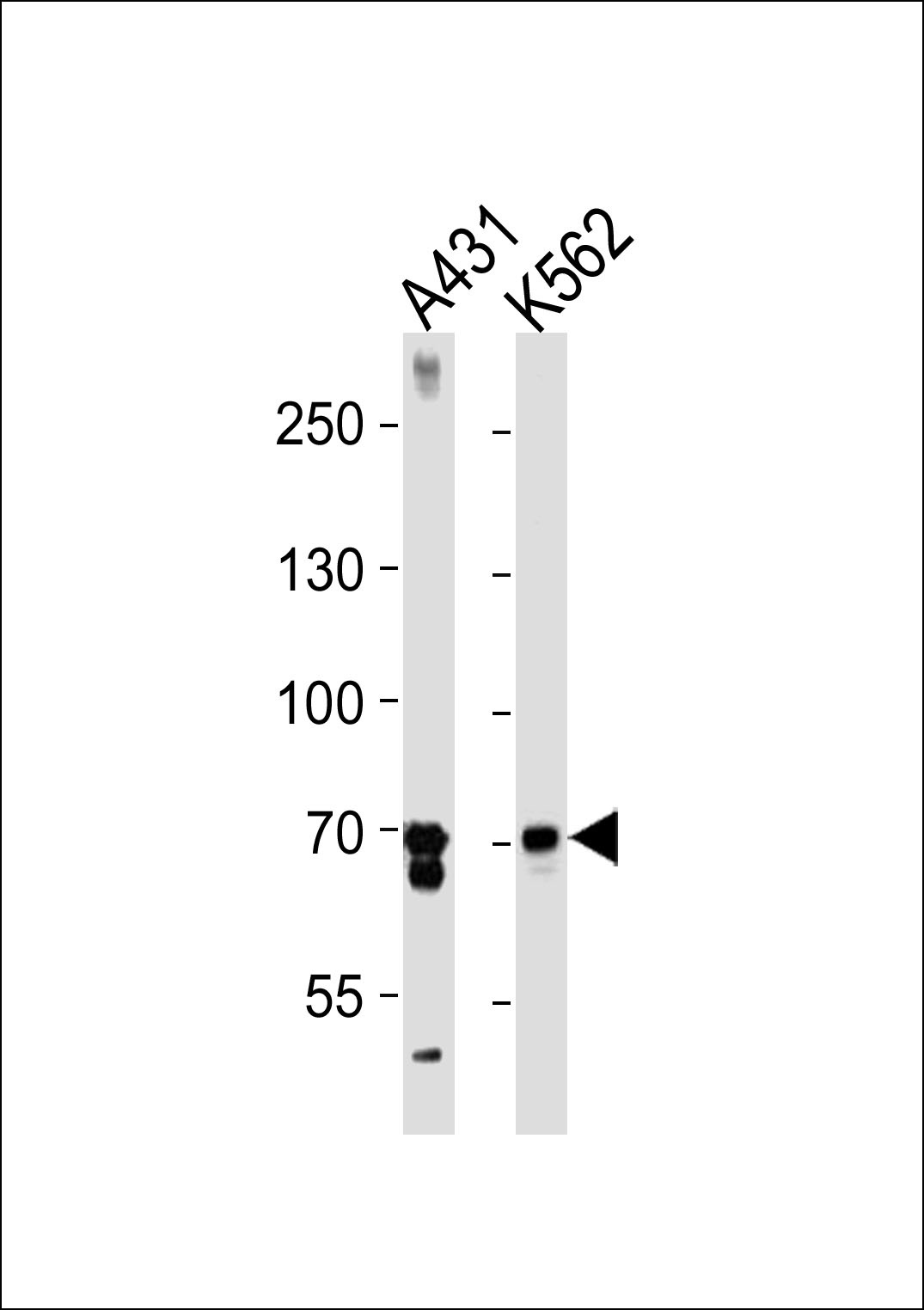
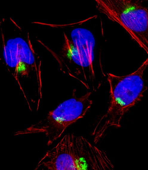
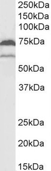
| Application notes | Peptide ELISA: antibody detection limit dilution 1:32000.Western Blot:Approx 60kDa and 70-75kDa bands observed in lysates of cell line U937 and approx 70-75kDa band observed in Mouse Kidney and Skeletal Muscle lysates (calculated MW of 64.5kDa according to Human NP_542158.1 and 68.7kDa Human NP_000953.2, and 69.0kDa according to Mouse NP_032995.1). The observed molecular weights correspond to earlier findings with different antibodies from other commercial sources. Recommended concentration: 0.5-2ug/ml.Immunofluorescence: Strong expression of the protein seen in the Golgi apparatus and vesicles of NIH3T3 cells. Recommended concentration: 5ug/ml. |
| Expiration Date | 12 months from date of receipt. |
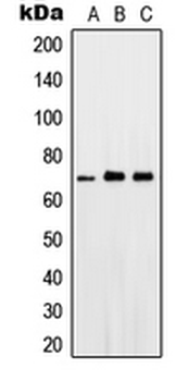
orb1247911 (2 ug/ml) staining of U937 lysate (35 ug protein in RIPA buffer). Primary incubation was 1 hour. Detected by chemiluminescence.
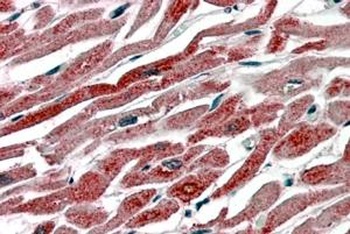
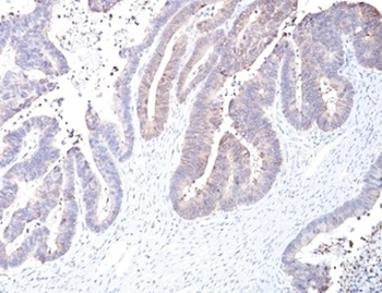
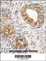
orb1247911 (3.8 ug/ml) staining of paraffin embedded Human Heart. Steamed antigen retrieval with citrate buffer pH 6, AP-staining. This data is from a previous batch, not on sale.
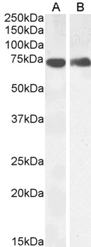
orb1247911 (0.5 ug/ml) staining of Mouse Kidney (A) and Skeletal Muscle (B) lysate (35 ug protein in RIPA buffer). Primary incubation was 1 hour. Detected by chemiluminescence.
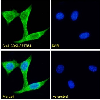
orb1247911 Immunofluorescence analysis of paraformaldehyde fixed NIH3T3 cells, permeabilized with 0.15% Triton. Primary incubation 1hr (5 ug/ml) followed by Alexa Fluor 488 secondary antibody (2 ug/ml), showing Golgi and vesicle staining.
- Item 1 of 7
Anti-COX1/Cyclooxygenase 1/PTGS1 Antibody [orb180700]
FC, IHC, WB
Human, Mouse, Rat
Rabbit
Polyclonal
Unconjugated
10 μg, 100 μg - Item 1 of 4
- Item 1 of 2
- Item 1 of 2
Cyclooxygenase 1 Rabbit Polyclonal Antibody [orb10448]
IF, IHC-Fr, IHC-P, WB
Bovine, Canine, Porcine, Rat, Sheep
Human, Mouse
Rabbit
Polyclonal
Unconjugated
50 μl, 100 μl, 200 μl - Item 1 of 3