You have no items in your shopping cart.
Cart summary
Item 1 of 10
Item 1 of 10
Collagen Type III Antibody
Catalog Number: orb345349

| Catalog Number | orb345349 |
|---|---|
| Category | Antibodies |
| Description | Collagen Type III antibody |
| Species/Host | Rabbit |
| Clonality | Polyclonal |
| Tested applications | DOT, ELISA, IHC, IP, WB |
| Reactivity | Bovine, Human, Porcine |
| Isotype | IgG |
| Immunogen | Collagen Type III from human and bovine placenta |
| Concentration | 1.16 mg/mL |
| Dilution range | ELISA: 1:5,000 - 1:50,000, IHC: 1:50 - 1:200, IP: 1:100, WB: 1:1,000 - 1:10,000 |
| Form/Appearance | Liquid (sterile filtered) |
| Purity | Collagen III Antibody has been prepared by immunoaffinity chromatography using immobilized antigens. Some class-specific anti-collagens may be specific for three-dimensional epitopes which may result in diminished reactivity with denatured collagen or formalin-fixed, paraffin embedded tissues. This antibody reacts with most mammalian Type III collagens and has expected cross-reactivity with Type I and negligible cross reactivity with Type II, IV, V or VI collagens. Non-specific cross-reaction of anti-collagen antibodies with other human serum proteins or non-collagen extracellular matrix proteins has not been tested. |
| Conjugation | Unconjugated |
| UniProt ID | P02461 |
| NCBI | NP_000081.1 |
| Storage | Store vial at 4° C prior to opening. This product is stable at 4° C as an undiluted liquid. Dilute only prior to immediate use. For extended storage, mix with an equal volume of glycerol, aliquot contents and freeze at -20° C or below. Avoid cycles of freezing and thawing. |
| Buffer/Preservatives | 0.01% (w/v) Sodium Azide |
| Alternative names | rabbit anti-Collagen Type III antibody, Collagen t Read more... |
| Note | For research use only |
| Application notes | Anti-Collagen Type III has been tested by dot Blot, western blot, and IHC and is useful for indirect trapping ELISA for quantitation of antigen in serum using a standard curve, immunoprecipitation, native (non-denaturing, non-dissociating) PAGE, immunohistochemistry, and western blotting for highly sensitive qualitative analysis. |
| Expiration Date | 12 months from date of receipt. |

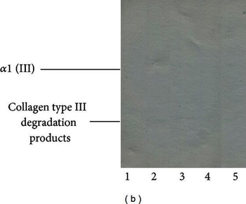
(a) Typical electropherogram of α (I) subunits, oligomers β/γ (I), and collagen type I degradation products, released from healthy and burnt skin. Collagenous components released from tissue samples using pepsin were submitted to 4–15% gradient SDS-PAGE, in nonreducing conditions. (b) Interferences of collagen type III, in electrophoretic profiles of collagen type I components extracted from healthy and burned skin. Collagen components were submitted to electrophoresis in the absence of dithiothreitol (reducing disulfide bonds) and subsequently—after electrotransfer to Immobilon—subjected to reaction with collagen type III antibodies. Lane 1: components of collagen type I isolated from healthy skin. Lane 2: components of collagen type I isolated from burned skin treated with propolis. Lane 3: components of collagen type I isolated from burned skin treated with AgSD. Lane 4: components of collagen type I isolated from burned skin treated with propolis vehicle. Lane 5: components of collagen type I isolated from burned skin treated with NaCl.

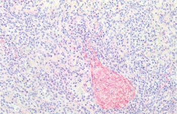
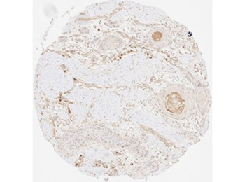
Immunohistochemistry of Rabbit Anti-Collagen Type III Antibody. Tissue: FFPE normal human spleen tissues (10X). Antigen Retrieval: 0.01 M sodium citrate buffer for 20 minutes. Primary Antibody: Anti-Collagen Type III at 5 µl/mL for 45 mins at RT. Staining: Anti-Rabbit biotinylated secondary antibody for 30 min at RT. Alkaline phosphatase streptavidin for 30 min at RT. Alkaline phosphatase chromogen substrate for 30 min at RT. The stained slides were evaluated by a pathologist to confirm staining specificity.

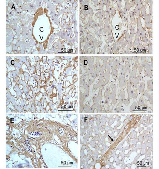
Immunohistochemistry of Rabbit Anti-collagen type III antibody. Tissue: right lobe of the liver section. A: Central Vein (CV) fibrosis, B: Non-fibrotic CV, C: Perisinusodial fibrosis, D: Non-fibrotic area, E: Protat tract fibrosis, F: Septal fibrosis (arrow). Fixation: formalin fixed paraffin embedded. Antigen retrieval: not required. Primary antibody: Anti-collagen type III at 1:500 for 4°C for 24hr. Secondary antibody: Peroxidase biotin-streptavidin rabbit secondary antibody at 1:10000 for 45 min at RT. Localization: Anti-collagen type III is intra and extracellular. Staining: 3.3'-diaminobenzidine tetrahydrochloride was used as the chromogen. Nuclei were counterstained purple with hematoxylin.
Immunohistochemistry results of Rabbit Anti-Collagen Type I Antibody. Tissue: human stroma in colorectal cancer. Fixation: FFPE. Antigen Retrieval: HIER using Tris-EDTA-citrate buffer pH7.8 for 5 min. Blocking: Peroxidase-Blocking Solution for 10 min. Primary Antibody: Anti-Collagen Type I (p/n orb345343) at 1:15 for 1 hr at 37 °C. Secondary Antibody: Dako REAL EnVision Detection Kit, Polymer-HRP, Rabbit/Mouse. Counterstain: Hematoxylin for 15 sec. Substrate: DAB-Chromogen, Rabbit/Mouse. Staining/Results: Fibrillar collagen III staining of the stroma in a colorectal cancer.
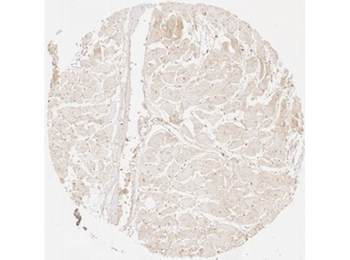
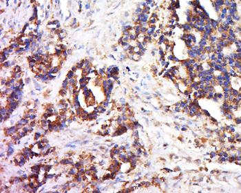
Immunohistochemistry results of Rabbit Anti-Collagen Type III Antibody. Tissue: human heart muscle. Fixation: FFPE. Antigen Retrieval: HIER using Tris-EDTA-citrate buffer pH7.8 for 5 min. Blocking: Peroxidase-Blocking Solution for 10 min. Primary Antibody: Anti-Collagen Type III (p/n orb345349) at 1:15 for 1 hr at 37 °C. Secondary Antibody: Dako REAL EnVision Detection Kit, Polymer-HRP, Rabbit/Mouse. Counterstain: Hematoxylin for 15 sec. Substrate: DAB-Chromogen, Rabbit/Mouse. Staining/Results: Distinct fibrillar collagen III staining surrounding each heart muscle cell.

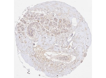
Immunohistochemistry results of Rabbit Anti-Collagen Type III Antibody. Tissue: human non-cancerous breast tissue. Fixation: FFPE. Antigen Retrieval: HIER using Tris-EDTA-citrate buffer pH7.8 for 5 min. Blocking: Peroxidase-Blocking Solution for 10 min. Primary Antibody: Anti-Collagen Type III (p/n orb345349) at 1:225 for 1 hr at 37 °C. Secondary Antibody: Dako REAL EnVision Detection Kit, Polymer-HRP, Rabbit/Mouse. Counterstain: Hematoxylin for 15 sec. Substrate: DAB-Chromogen, Rabbit/Mouse. Staining/Results: Fibrillar collagen III staining in non-cancerous breast tissue showing considerable sclerosis.
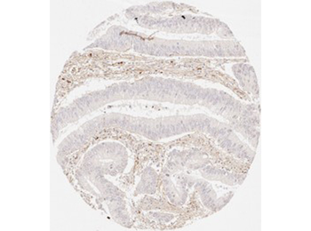
Immunohistochemistry results of Rabbit Anti-Collagen Type III Antibody. Tissue: human oral cavity. Fixation: FFPE. Antigen Retrieval: HIER using Tris-EDTA-citrate buffer pH7.8 for 5 min. Blocking: Peroxidase-Blocking Solution for 10 min. Primary Antibody: Anti-Collagen Type III (p/n orb345349) at 1:15 for 1 hr at 37 °C. Secondary Antibody: Dako REAL EnVision Detection Kit, Polymer-HRP, Rabbit/Mouse. Counterstain: Hematoxylin for 15 sec. Substrate: DAB-Chromogen, Rabbit/Mouse. Staining/Results: Distinct fibrillar collagen III staining of the stroma in squamous cell carcinoma of the oral cavity.

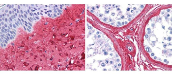
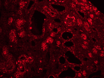
Biorbyt anti collagen III antibody (1:400, 45 min RT) showed strong staining in FFPE sections of human skin(left, dermis) with moderate to strong red staining and testis (right) where strong staining was observed within connective tissue between seminiferous tubules. The antibody showed strong extracellular staining within connective tissues across many organs with minimal background staining. Slides were steamed in 0.01 M sodium citrate buffer, pH6.0 at 99-100°C - 20 minutes for antigen retrieval.
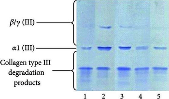
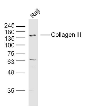
Typical electropherogram of α (III) subunits, oligomers β/γ (III), and collagen type III degradation products, released from healthy and burned skin. Collagenous components released from tissue samples using pepsin were submitted to 4–15% gradient SDS-PAGE, western blotted, and probed with collagen type III antibodies. Lane 1: components of collagen type I isolated from healthy skin. Lane 2: components of collagen type I isolated from burned skin treated with propolis. Lane 3: components of collagen type I isolated from burned skin treated with AgSD. Lane 4: components of collagen type I isolated from burned skin treated with propolis vehicle. Lane 5: components of collagen type I isolated from burned skin treated with NaCl.
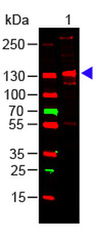
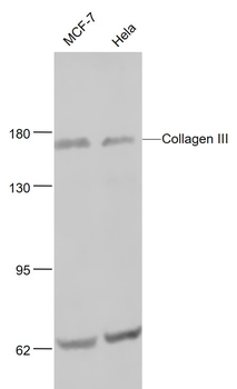
Western Blot of Rabbit Anti-COLLAGEN III Antibody. Lane 1: Human Collagen III (p/n orb750383). Load: 100 ng per lane. Primary antibody: Collagen III Antibody at 1:1000 o/n at 4°C. Secondary antibody: DyLight™ 649 Goat anti-rabbit (p/n orb347673) at 1:20000 for 30 min at RT. Block: orb348637 for 30 min at RT. Predicted/Observed size: 138 kDa, 138 kDa.
- Item 1 of 10
Collagen Type III Antibody [orb345350]
DOT, ELISA, IHC, IP, WB
Bovine, Human, Porcine
Rabbit
Polyclonal
Unconjugated
500 μg - Item 1 of 6
Collagen III antibody [orb304041]
ELISA, IHC-P
Bovine, Canine, Gallus, Human, Mouse, Rat
Rabbit
Polyclonal
Unconjugated
100 μg - Item 1 of 3
Collagen III Rabbit Polyclonal Antibody [orb10437]
IF, IHC-Fr, IHC-P, WB
Bovine, Gallus, Mouse, Rat
Canine, Human, Rabbit
Rabbit
Polyclonal
Unconjugated
200 μl, 50 μl, 100 μl - Item 1 of 3
Anti-Collagen 3 alpha 1 Antibody [orb213759]
IF, IH, WB
Human, Mouse, Porcine, Rabbit, Rat
Rabbit
Polyclonal
Unconjugated
30 μl, 100 μl, 200 μl - Item 1 of 3
Collagen III Rabbit Polyclonal Antibody [orb10438]
ELISA, IF, IHC-Fr, IHC-P, WB
Bovine, Mouse
Human, Rat
Rabbit
Polyclonal
Unconjugated
50 μl, 100 μl, 200 μl