You have no items in your shopping cart.
Cart summary
Item 1 of 5
Item 1 of 5
COL1A1 antibody
Catalog Number: orb345868

| Catalog Number | orb345868 |
|---|---|
| Category | Antibodies |
| Description | COL1A1 antibody |
| Species/Host | Rabbit |
| Clonality | Polyclonal |
| Tested applications | DOT, ELISA, FC, IHC, IP, Multiplex Assay, WB |
| Reactivity | Human, Mouse, Rat |
| Isotype | IgG |
| Immunogen | Collagen Type I from human and bovine placenta. |
| Concentration | 1.0 mg/mL |
| Dilution range | ELISA: 1:3,000 - 1:6,000, FC: User Optimized, IHC: 1:50 - 1:200, IP: 1:100, WB: 1:3,000 - 1:6,000 |
| Form/Appearance | Lyophilized |
| Purity | This product has been prepared by immunoaffinity chromatography using immobilized antigens. Some class-specific anti-collagens may be specific for three-dimensional epitopes which may result in diminished reactivity with denatured collagen or formalin-fixed, paraffin embedded tissues. This antibody reacts with most mammalian Type I collagens and has expected cross-reactivity with Type III and negligible cross reactivity with Type II, IV, V or VI collagens. Non-specific cross-reaction of anti-collagen antibodies with other human serum proteins or non-collagen extracellular matrix proteins has not been tested. |
| Conjugation | Biotin |
| UniProt ID | P02452 |
| NCBI | NP_000079.2 |
| Storage | Store vial at 4° C prior to restoration. Restore with 0.1 mL of deionized water (or equivalent). For extended storage aliquot contents and freeze at -20° C or below. Avoid cycles of freezing and thawing. Centrifuge product if not completely clear after standing at room temperature. This product is stable for several weeks at 4° C as an undiluted liquid. Dilute only prior to immediate use. Expiration date is one (1) year from date of restoration. |
| Buffer/Preservatives | 0.01% (w/v) Sodium Azide |
| Alternative names | rabbit anti-collagen type I antibody biotin conjug Read more... |
| Note | For research use only |
| Application notes | Anti-COLLAGEN Type I Antibody Biotin Conjugated has been tested by dot blot and Flow Cytometry and is suitable for western blot, immunoprecipitation, Flow Cytometry, and immunohistochemistry. Researchers should determine optimal titers for applications that are not stated below. |
| Expiration Date | 12 months from date of receipt. |
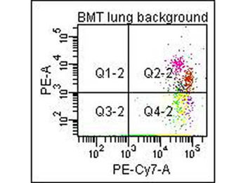
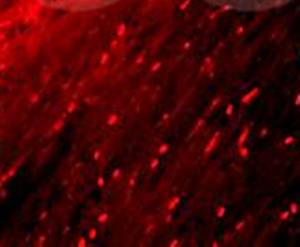
Flow Cytometry of Anti-Collagen Type I Biotin Conjugated Antibody (orb345868). Cells: mouse lung. Stimulation: none. Primary antibody: biotin conjugated anti-collagen type I antibody. Secondary antibody: PE-conjugated CD45 and PE-conjugated anti-collagen type I secondary antibody.
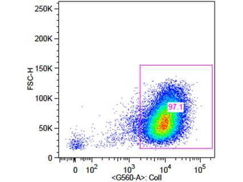
Flow Cytometry of Rabbit Anti-Collagen 1 Antibody. Cells: primary adult human dermal fibroblast cells. Stimulation: none. Primary antibody: Biotin-Conjugated Collagen 1 antibody (orb345868) at 5 µg/mL for 45 min at 4°C. Secondary antibody: Rabbit Streptavidin, R-PE antibody at 1:500 for 15 min at RT.
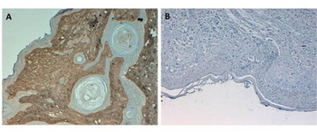
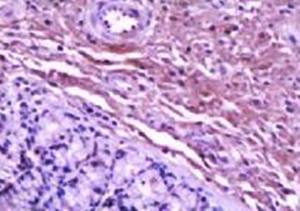
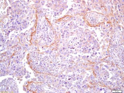
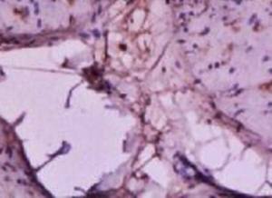
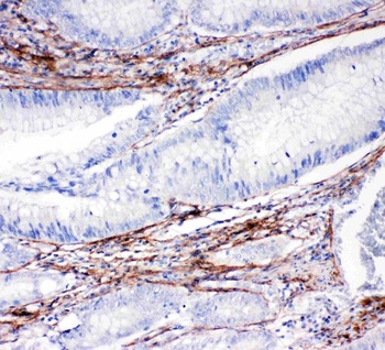
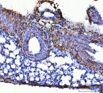
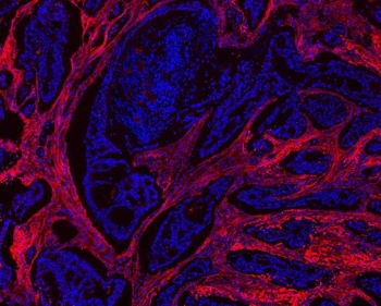
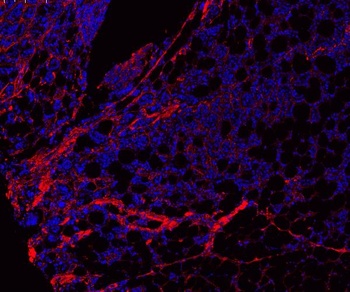
Immunohistochemistry of Rabbit Anti-Collagen Type I Antibody. Tissue: Human Skin at pH6. Fixation: formalin fixed paraffin embedded. Antigen retrieval: not required. Primary antibody: Collagen Type I antibody at 10 µg/mL for 1 h at RT. Secondary antibody: Peroxidase rabbit secondary antibody at 1:10000 for 45 min at RT. Localization: Collagen Type I is secreted in the extracellular matrix. Staining: Collagen Type I as precipitated brown signal (A) with hematoxylin purple nuclear counterstain. With corresponding negative conrol (B).
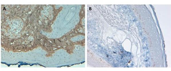
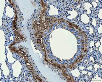
Immunohistochemistry of Rabbit Anti-Collagen Type I Antibody. Tissue: Human Skin at pH9. Fixation: formalin fixed paraffin embedded. Antigen retrieval: not required. Primary antibody: Collagen Type I antibody at 10 µg/mL for 1 h at RT. Secondary antibody: Peroxidase rabbit secondary antibody at 1:10000 for 45 min at RT. Localization: Collagen Type I is secreted in the extracellular matrix. Staining: Collagen Type I as precipitated brown signal (A) with hematoxylin purple nuclear counterstain. With corresponding negative conrol (B).
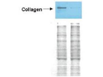
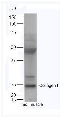
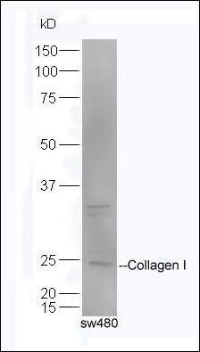
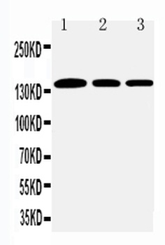
Western Blot of Rabbit anti-Collagen I antibody. Lane 1: Wistar rat hepatic stellate cells (HSC) in control (GFP-transduced). Lane 2: PPARg-transduced cell lysates. Load: 100 µg per lane. Protein staining shown below western blot depicts equal protein loading. Primary antibody: Anti-Collagen I antibody at 0.2–2 µg/10 ml for overnight at 4°C. Secondary antibody: horseradish peroxidase-conjugated rabbit secondary antibody at 1 µg/10 ml for overnight at 4°C. Block: TBS with 5% Non-fat milk. Predicted/Observed size: 138.9 kDa for Collagen I. Other band(s): none.
- Item 1 of 9
- Item 1 of 6
Collagen I antibody [orb312178]
IHC-P, WB
Bovine, Equine, Monkey, Sheep
Canine, Human, Mouse, Rabbit, Rat
Rabbit
Polyclonal
Unconjugated
100 μl, 50 μl, 200 μl - Item 1 of 8
COL1A1 antibody [orb1294293]
IF, IHC, WB
Human, Mouse, Rabbit
Rabbit
Polyclonal
Unconjugated
100 μl, 25 μl - Item 1 of 8
Collagen I/COL1A1 Antibody [orb107158]
ICC, IF, IHC, WB
Hamster
Human, Mouse, Rat
Rabbit
Polyclonal
Unconjugated
10 μg, 100 μg - Item 1 of 6


Submit a review
Filter by Rating

- 5 stars

- 4 stars
- 3 stars
- 2 stars
- 1 stars
