You have no items in your shopping cart.
Cart summary
Item 1 of 5
Item 1 of 5
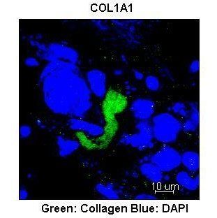
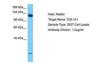
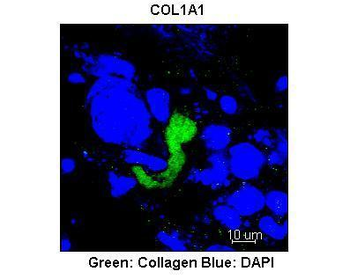
COL1A1 antibody
Catalog Number: orb345844

| Catalog Number | orb345844 |
|---|---|
| Category | Antibodies |
| Description | COL1A1 antibody |
| Species/Host | Rabbit |
| Clonality | Polyclonal |
| Tested applications | ELISA, FC, IHC, IP, WB |
| Reactivity | Bovine, Human, Mouse, Rat |
| Isotype | IgG |
| Immunogen | Collagen Type I from human and bovine placenta. |
| Concentration | 1.0 mg/mL |
| Dilution range | ELISA: 1:3,000 - 1:6,000, FC: 5µg/mL, IHC: 1:50 - 1:200, IP: 1:100, WB: 1:3,000 - 1:6,000 |
| Form/Appearance | Lyophilized |
| Purity | This product has been prepared by immunoaffinity chromatography using immobilized antigens. Some class-specific anti-collagens may be specific for three-dimensional epitopes which may result in diminished reactivity with denatured collagen or formalin-fixed, paraffin embedded tissues. This antibody reacts with most mammalian Type I collagens and has expected cross-reactivity with Type III and negligible cross reactivity with Type II, IV, V or VI collagens. Non-specific cross-reaction of anti-collagen antibodies with other human serum proteins or non-collagen extracellular matrix proteins has not been tested. |
| Conjugation | HRP |
| UniProt ID | P02452 |
| NCBI | NP_000079.2 |
| Storage | Store vial at 4° C prior to restoration. Restore with 0.05 mL of deionized water (or equivalent). For extended storage aliquot contents and freeze at -20° C or below. Avoid cycles of freezing and thawing. Centrifuge product if not completely clear after standing at room temperature. This product is stable for several weeks at 4° C as an undiluted liquid. Dilute only prior to immediate use. Expiration date is one (1) year from date of restoration. |
| Buffer/Preservatives | 0.01% (w/v) Thimerosal |
| Alternative names | rabbit anti-collagen type I antibody peroxidase co Read more... |
| Note | For research use only |
| Application notes | Anti-COLLAGEN Type I Peroxidase Conjugated Antibody is suitable for western blot, immunoprecipitation, Flow Cytometry, and immunohistochemistry. Researchers should determine optimal titers for applications that are not stated below. |
| Expiration Date | 12 months from date of receipt. |
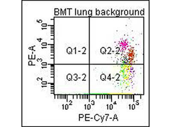
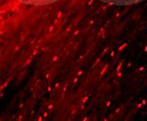
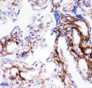
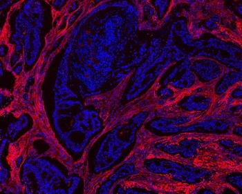
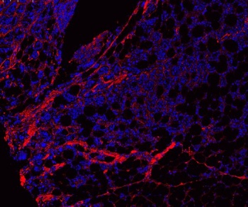
Flow Cytometry of Anti-Collagen Type I Biotin Conjugated Antibody (orb345868). Cells: mouse lung. Stimulation: none. Primary antibody: biotin conjugated anti-collagen type I antibody. Secondary antibody: PE-conjugated CD45 and PE-conjugated anti-collagen type I secondary antibody.
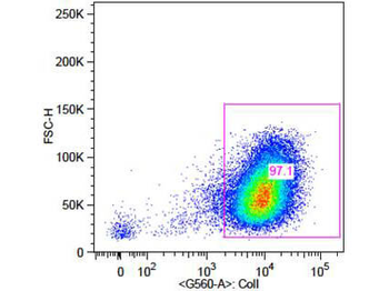
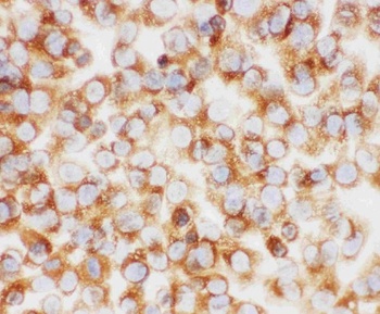
Flow Cytometry of Rabbit Anti-Collagen 1 Antibody. Cells: primary adult human dermal fibroblast cells. Stimulation: none. Primary antibody: Biotin-Conjugated Collagen 1 antibody (orb345868) at 5 µg/mL for 45 min at 4°C. Secondary antibody: Rabbit Streptavidin, R-PE antibody at 1:500 for 15 min at RT.
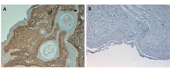
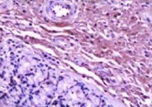
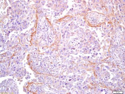
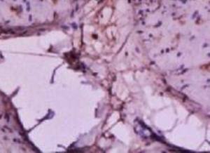
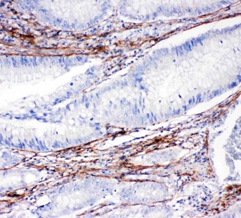
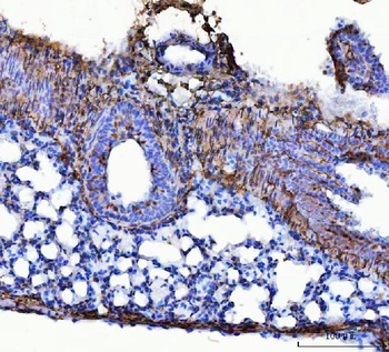
Immunohistochemistry of Rabbit Anti-Collagen Type I Antibody. Tissue: Human Skin at pH6. Fixation: formalin fixed paraffin embedded. Antigen retrieval: not required. Primary antibody: Collagen Type I antibody at 10 µg/mL for 1 h at RT. Secondary antibody: Peroxidase rabbit secondary antibody at 1:10000 for 45 min at RT. Localization: Collagen Type I is secreted in the extracellular matrix. Staining: Collagen Type I as precipitated brown signal (A) with hematoxylin purple nuclear counterstain. With corresponding negative conrol (B).
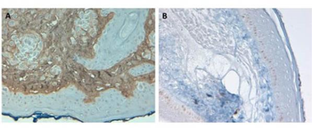
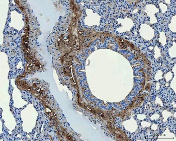
Immunohistochemistry of Rabbit Anti-Collagen Type I Antibody. Tissue: Human Skin at pH9. Fixation: formalin fixed paraffin embedded. Antigen retrieval: not required. Primary antibody: Collagen Type I antibody at 10 µg/mL for 1 h at RT. Secondary antibody: Peroxidase rabbit secondary antibody at 1:10000 for 45 min at RT. Localization: Collagen Type I is secreted in the extracellular matrix. Staining: Collagen Type I as precipitated brown signal (A) with hematoxylin purple nuclear counterstain. With corresponding negative conrol (B).
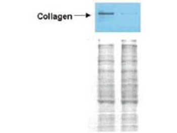
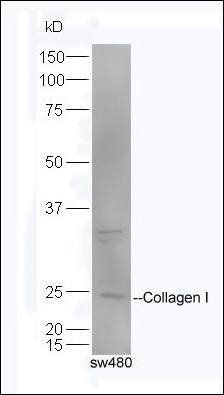
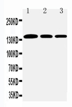
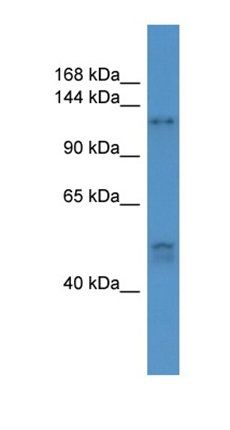
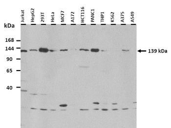
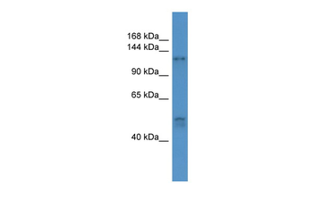
Western Blot of Rabbit anti-Collagen I antibody. Lane 1: Wistar rat hepatic stellate cells (HSC) in control (GFP-transduced). Lane 2: PPARg-transduced cell lysates. Load: 100 µg per lane. Protein staining shown below western blot depicts equal protein loading. Primary antibody: Anti-Collagen I antibody at 0.2–2 µg/10 ml for overnight at 4°C. Secondary antibody: horseradish peroxidase-conjugated rabbit secondary antibody at 1 µg/10 ml for overnight at 4°C. Block: TBS with 5% Non-fat milk. Predicted/Observed size: 138.9 kDa for Collagen I. Other band(s): none.
- Item 1 of 9
- Item 1 of 6
Collagen I antibody [orb312178]
IHC-P, WB
Bovine, Equine, Monkey, Sheep
Canine, Human, Mouse, Rabbit, Rat
Rabbit
Polyclonal
Unconjugated
100 μl, 50 μl, 200 μl - Item 1 of 8
COL1A1 antibody [orb1294293]
IF, IHC, WB
Human, Mouse, Rabbit
Rabbit
Polyclonal
Unconjugated
100 μl, 25 μl - Item 1 of 8
Collagen I/COL1A1 Antibody [orb107158]
ICC, IF, IHC, WB
Hamster
Human, Mouse, Rat
Rabbit
Polyclonal
Unconjugated
10 μg, 100 μg - Item 1 of 7
Collagen I antibody [orb331081]
IHC, WB
Bovine, Canine, Equine, Guinea pig, Rabbit, Rat
Human, Mouse
Rabbit
Polyclonal
Unconjugated
100 μl


Submit a review
Filter by Rating

- 5 stars

- 4 stars
- 3 stars
- 2 stars
- 1 stars
