You have no items in your shopping cart.
Cart summary
Item 1 of 5
Item 1 of 5
CLU Antibody (N-term)
Catalog Number: orb1938586

| Catalog Number | orb1938586 |
|---|---|
| Category | Antibodies |
| Description | Affinity Purified Rabbit Polyclonal Antibody (Pab) |
| Species/Host | Rabbit |
| Clonality | Polyclonal |
| Clone Number | RB18701 |
| Tested applications | FC, IHC-P, WB |
| Reactivity | Human, Mouse, Rat |
| Isotype | Rabbit IgG |
| Dilution range | WB: 1:1000, WB: 1:500-1000, WB: 1:500-1000, IHC-P-Leica: 1:500, FC: 1:25 |
| Form/Appearance | Purified polyclonal antibody supplied in PBS with 0.09% (W/V) sodium azide. This antibody is purified through a protein A column, followed by peptide affinity purification. |
| Conjugation | Unconjugated |
| MW | 52495 Da |
| Target | This CLU antibody is generated from rabbits immunized with a KLH conjugated synthetic peptide between 71-99 amino acids from the N-terminal region of human CLU. |
| UniProt ID | P10909 |
| NCBI | NP_001822.3 |
| Storage | Maintain refrigerated at 2-8°C for up to 2 weeks. For long term storage store at -20°C in small aliquots to prevent freeze-thaw cycles |
| Alternative names | Clusterin, Aging-associated gene 4 protein, Apolip Read more... |
| Note | For research use only |
| Expiration Date | 12 months from date of receipt. |

All lanes: Anti-CLU Antibody (N-term) at 1:1000 dilution. Lane 1: HepG2 whole cell lysate. Lane 2: MCF-7 whole cell lysate. Lane 3: Caco2 whole cell lysate. Lane 4: Hela whole cell lysate. Lane 5: A549 whole cell lysate. Lysates/proteins at 20 µg per lane. Secondary Goat Anti-Rabbit IgG, (H+L), Peroxidase conjugated at 1/10000 dilution. Predicted band size: 52 kDa. Blocking/Dilution buffer: 5% NFDM/TBST.
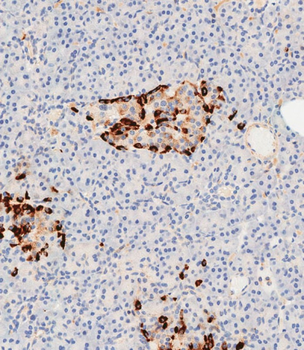
Immunohistochemical analysis of paraffin-embedded human pancreas tissue performed on the Leica BOND RXm. Samples were incubated with primary antibody (1/500) for 1 hours at room temperature. A undiluted biotinylated CRF Anti-Polyvalent HRP Polymer antibody was used as the secondary Antibody.
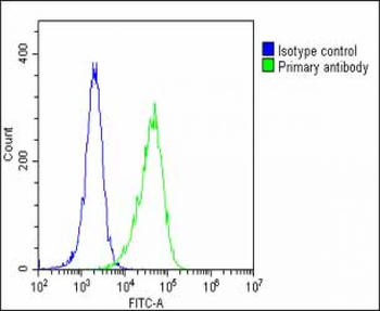
Overlay histogram showing Hela cells (green line). The cells were fixed with 2% paraformaldehyde (10 min) and then permeabilized with 90% methanol for 10 min. The cells were then icubated in 2% bovine serum albumin to block non-specific protein-protein interactions followed by the antibody (1:25 dilution) for 60 min at 37°C. The secondary antibody used was Goat-Anti-Rabbit IgG, DyLight 488 Conjugated Highly Cross-Adsorbed (1583138) at 1/200 dilution for 40 min at 37°C. Isotype control antibody (blue line) was rabbit IgG1 (1 μg/1x10^6 cells) used under the same conditions. Acquisition of > 10000 events was performed.
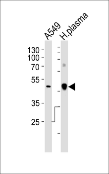
Western blot analysis of lysates from A549 cell line and human plasma tissue lysate (from left to right), using CLU Antibody (N-term). diluted at 1:1000 at each lane. A goat anti-rabbit IgG H&L (HRP) at 1:10000 dilution was used as the secondary Antibody. Lysates at 35 ug per lane.
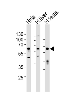
Western blot analysis of lysates from Hela cell line, human liver and human testis tissue lysate (from left to right), using CLU Antibody (N-term). diluted at 1:1000 at each lane. A goat anti-rabbit IgG H&L (HRP) at 1:10000 dilution was used as the secondary Antibody. Lysates at 35 ug per lane.
