You have no items in your shopping cart.
Cart summary
Item 1 of 7
Item 1 of 7
Cleaved LC3A Antibody
Catalog Number: orb1933527

| Catalog Number | orb1933527 |
|---|---|
| Category | Antibodies |
| Description | Affinity Purified Rabbit Polyclonal Antibody (Pab) |
| Species/Host | Rabbit |
| Clonality | Polyclonal |
| Clone Number | RB38908 |
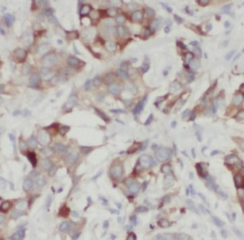
| Tested applications | ICC, IF, IHC-P, WB |
| Predicted Reactivity | Rat, Zebrafish |
| Reactivity | Human, Mouse |
| Isotype | Rabbit IgG |
| Dilution range | IF: 1:25, IF: 1:25, WB: 1:500, WB: 1:500, WB: 1:500, WB: 1:500, WB: 1:1000 |
| Form/Appearance | Purified polyclonal antibody supplied in PBS with 0.09% (W/V) sodium azide. This antibody is purified through a protein A column, followed by peptide affinity purification. |
| Conjugation | Unconjugated |
| Target | This Cleaved LC3A antibody is generated from rabbits immunized with a KLH conjugated synthetic peptide between 89-120 amino acids from human Cleaved LC3A or LC3B. |
| UniProt ID | Q9H492, Q9GZQ8 |
| NCBI | NP_852610.1, NP_115903.1 |
| Storage | Maintain refrigerated at 2-8°C for up to 2 weeks. For long term storage store at -20°C in small aliquots to prevent freeze-thaw cycles |
| Alternative names | Microtubule-associated proteins 1A/1B light chain Read more... |
| Note | For research use only |
| Expiration Date | 12 months from date of receipt. |

Western blot analysis of anti-cleaved-LC3 (APG8a) Pab in mouse brain tissue lysate. Cleaved-LC3 (APG8a) was detected using the purified Pab.
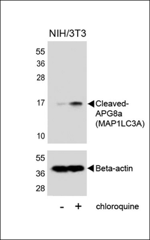
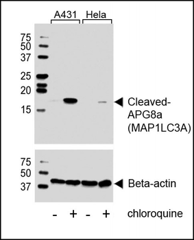
Western blot analysis of lysates from NIH/3T3 cells, untreated or treated with chloroquine, using Cleaved-APG8a (MAP1LC3A) (upper) or Beta-actin (lower).
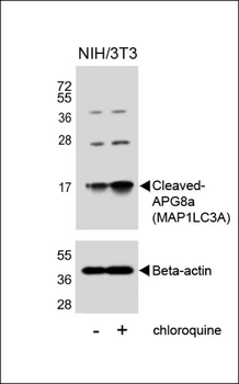
Western blot analysis of lysates from NIH/3T3 cells, untreated or treated with chloroquine, using Cleaved-APG8a (MAP1LC3A) (upper) or Beta-actin (lower).
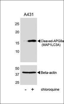
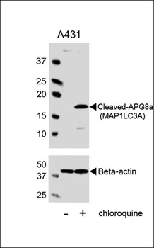
Western blot analysis of lysates from A431 cell line, untreated or treated with chloroquine, 100ng/ml, using Cleaved-APG8a (MAP1LC3A) (upper) or Beta-actin (lower).

Western blot analysis of lysates from A431 cell line, untreated or treated with chloroquine, 100ng/ml, using Cleaved-APG8a (MAP1LC3A) Antibody (upper) or Beta-actin (lower).

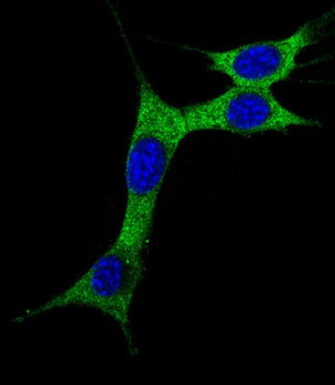
Immunofluorescent analysis of 4% paraformaldehyde-fixed, 0.1% Triton X-100 permeabilized NIH/3T3 (Mouse mouse embryonic fibroblasts cell line) cells labeling MAP1LC3A at 1/25 dilution, followed by Alexa Fluor 488-conjugated goat anti-rabbit IgG secondary antibody at 1/400 dilution (green). The nuclear counter stain is DAPI (blue). Immunofluorescence image showing cytoplasm on NIH/3T3 cell line.
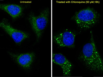
Immunofluorescent analysis of 4% paraformaldehyde-fixed, 0.1% Triton X-100 permeabilized Hela (Human Cervical epithelial adenocarcinoma cell line) cells labeling MAP1LC3A at 1/25 dilution, followed by Alexa Fluor 488-conjugated goat anti-rabbit IgG secondary antibody at 1/400 dilution (green). Immunofluorescence image showing vesicles staining on Hela cell line.The nuclear counter stain is DAPI (blue). The right image is Hela cells treated with Chloroquine 50μM for 16 h.
- Item 1 of 4
- Item 1 of 2

Cleaved LC3A Antibody [orb1167662]
ICC, IF, IHC-P, WB
Human, Mouse
Rabbit
Polyclonal
Unconjugated
100 μl, 30 μl