You have no items in your shopping cart.
Cart summary
Item 1 of 14
Item 1 of 14

| Catalog Number | orb10410 |
|---|---|
| Category | Antibodies |
| Description | CK7 Rabbit Polyclonal Antibody |
| Species/Host | Rabbit |
| Clonality | Polyclonal |
| Tested applications | FC, IF, IHC-Fr, IHC-P, WB |
| Predicted Reactivity | Mouse, Rat |
| Reactivity | Human, Mouse, Rat |
| Isotype | IgG |
| Immunogen | KLH conjugated synthetic peptide derived from the middle of human CK7 (251-350/469aa) |
| Concentration | 1mg/ml |
| Dilution range | WB=1:500-2000, IHC-P=1:100-500, IHC-F=1:100-500, IF=1:100-500, Flow-Cyt=0.2µg/Test |
| Form/Appearance | Liquid |
| Conjugation | Unconjugated |
| MW | 54 kDa |
| Target | KRT7 |
| UniProt ID | P08729 |
| RRID | AB_10746291 |
| Storage | Maintain refrigerated at 2-8°C for up to 2 weeks. For long term storage store at -20°C in small aliquots to prevent freeze-thaw cycles. |
| Buffer/Preservatives | 0.01M TBS (pH7.4) with 1% rAlbumin, 0.02% Proclin300 and 50% Glycerol. |
| Alternative names | Cytokeratin 7; Cytokeratin-7; CK 7; CK-7; K7; Kera Read more... |
| Note | For research use only |
| Expiration Date | 12 months from date of receipt. |
Caucheteur, Christophe et al. Immunosensing with Near-Infrared Plasmonic Optical Fiber Gratings Methods Mol Biol, 1571, 47-71 (2017)

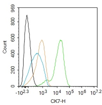
Blank control: A549. Primary Antibody (green line): Rabbit Anti-CK7 antibody (orb10410), Dilution: 1 µg/10^6 cells, Isotype Control Antibody (orange line): Rabbit IgG. Secondary Antibody: Goat anti-rabbit IgG-PE, Dilution: 0.2 µg/Test. Protocol, The cells were fixed with 4% PFA (10 min at room temperature) and then permeabilized with 20% PBST for 20 min at room temperature. The cells were then incubated in 5% BSA to block non-specific protein-protein interactions for 30 min at at room temperature. Cells stained with Primary Antibody for 30 min at room temperature. The secondary antibody used for 40 min at room temperature. Acquisition of 20000 events was performed.
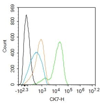
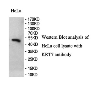
Blank control: Hela. Primary Antibody (green line): Rabbit Anti-CK7 antibody (orb10410), Dilution: 1 ug/Test, Secondary Antibody: Goat anti-rabbit IgG-FITC, Dilution: 0.5 ug/Test. Protocol, The cells were fixed with 4% PFA (10 min at room temperature) and then permeabilized with 90% ice-cold methanol for 20 min at -20°C. The cells were then incubated in 5% BSA to block non-specific protein-protein interactions for 30 min at room temperature. Cells stained with Primary Antibody for 30 min at room temperature. The secondary antibody used for 40 min at room temperature. Acquisition of 20000 events was performed.
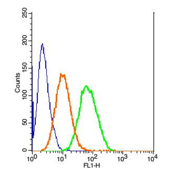
Blank control: Hepg2 (blue), Isotype Control Antibody: Rabbit IgG-FITC (orange), Primary Antibody Dilution: 1 µl in 100 µl 1X PBS containing 0.5% BSA (green).
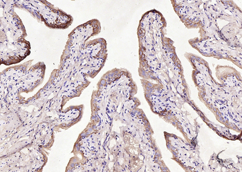
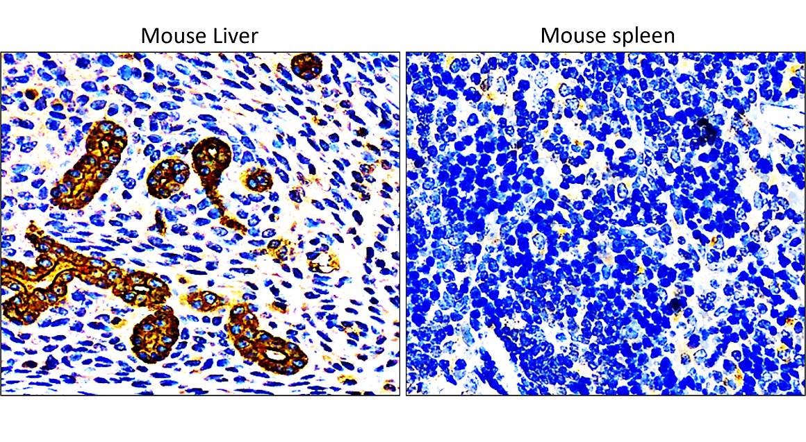
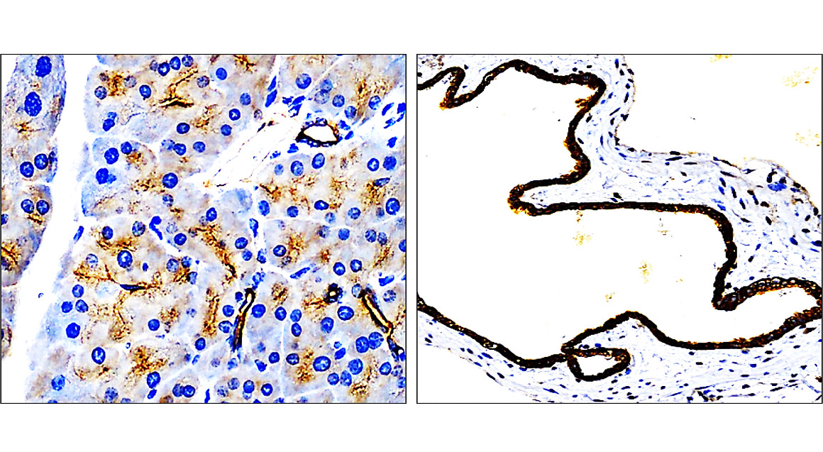
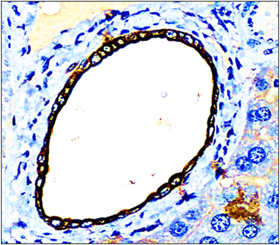
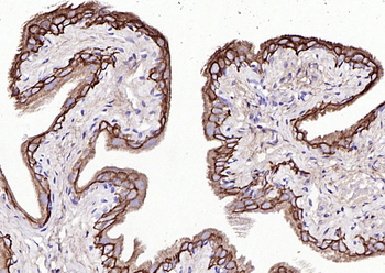
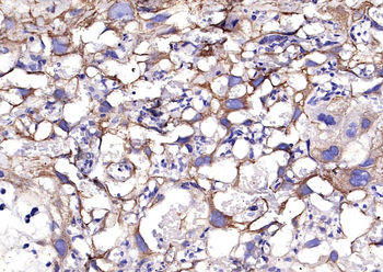
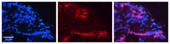
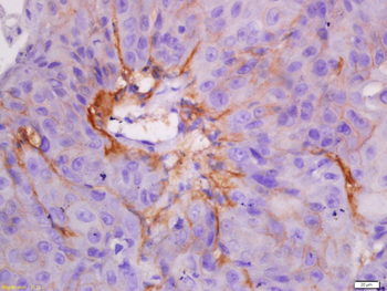
Paraformaldehyde-fixed, paraffin embedded (Rat bladder), Antigen retrieval by boiling in sodium citrate buffer (pH6.0) for 15 min, Block endogenous peroxidase by 3% hydrogen peroxide for 20 minutes, Blocking buffer (normal goat serum) at 37°C for 30 min, Antibody incubation with (CK7) Polyclonal Antibody, Unconjugated (orb10410) at 1:200 overnight at 4°C, followed by operating according to SP Kit (Rabbit) instructionsand DAB staining.
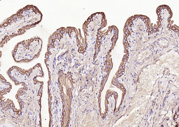
Paraformaldehyde-fixed, paraffin embedded (Rat bladder), Antigen retrieval by boiling in sodium citrate buffer (pH6.0) for 15 min, Block endogenous peroxidase by 3% hydrogen peroxide for 20 minutes, Blocking buffer (normal goat serum) at 37°C for 30 min, Antibody incubation with (CK7) Polyclonal Antibody, Unconjugated (orb10410) at 1:200 overnight at 4°C, followed by operating according to SP Kit (Rabbit) instructionsand DAB staining.
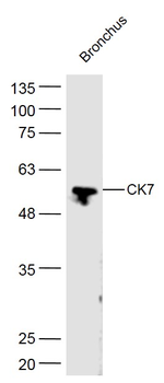
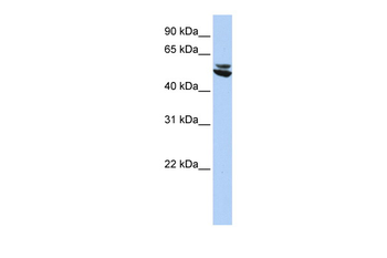
Sample: Bronchus (Mouse) Lysate at 40 ug, Primary: Anti-CK7 (orb10410) at 1/300 dilution, Secondary: IRDye800CW Goat Anti-Rabbit IgG at 1/20000 dilution, Predicted band size: 54 kD, Observed band size: 54 kD.
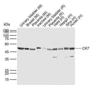
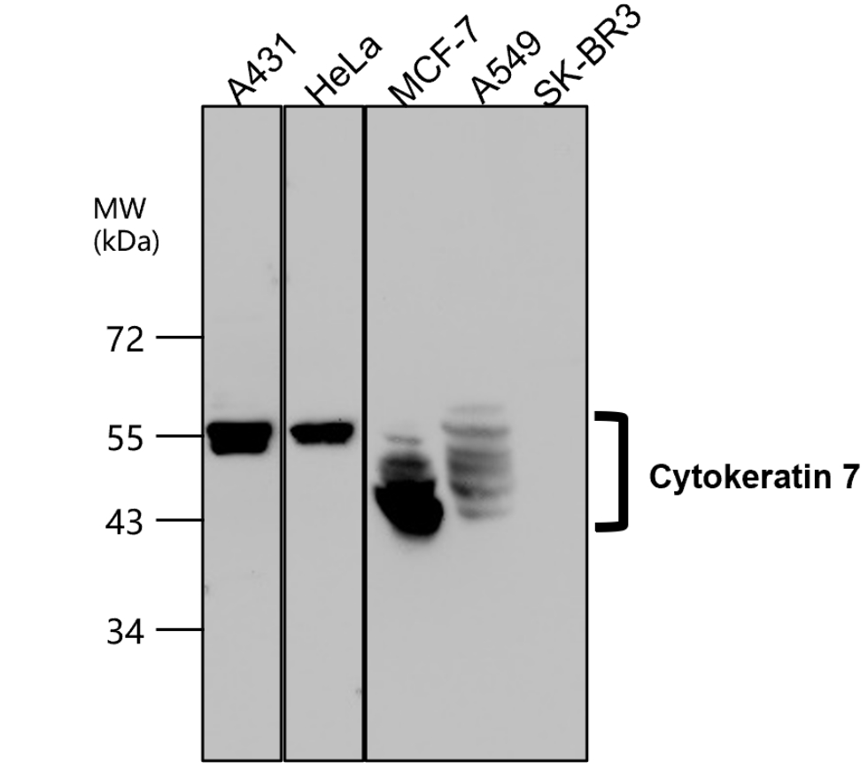
Sample: Lane 1: Mouse Urinary bladder tissue lysates, Lane 2: Mouse Breast tissue lysates, Lane 3: Mouse Placenta tissue lysates, Lane 4: Mouse trachea tissue lysates, Lane 5: Rat Urinary bladder tissue lysates, Lane 6: Rat Placenta tissue lysates, Lane 7: Human Hela cell lysates, Lane 8: Human Hepg2 cell lysates, Lane 9: Human Siha cell lysates, Lane 10: Human Huvec cell lysates, Primary: Anti-CK7 (orb10410) at 1/1000 dilution, Secondary: IRDye800CW Goat Anti-Rabbit IgG at 1/20000 dilution, Predicted band size: 54 kDa, Observed band size: 54 kDa.
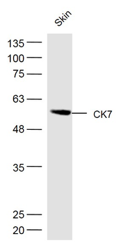
Sample: Skin (Mouse) Lysate at 40 ug, Primary: Anti-CK7 (orb10410) at 1/300 dilution, Secondary: IRDye800CW Goat Anti-Rabbit IgG at 1/20000 dilution, Predicted band size: 54 kD, Observed band size: 54 kD.

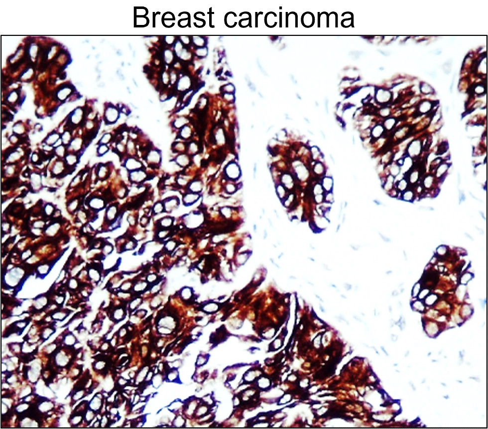
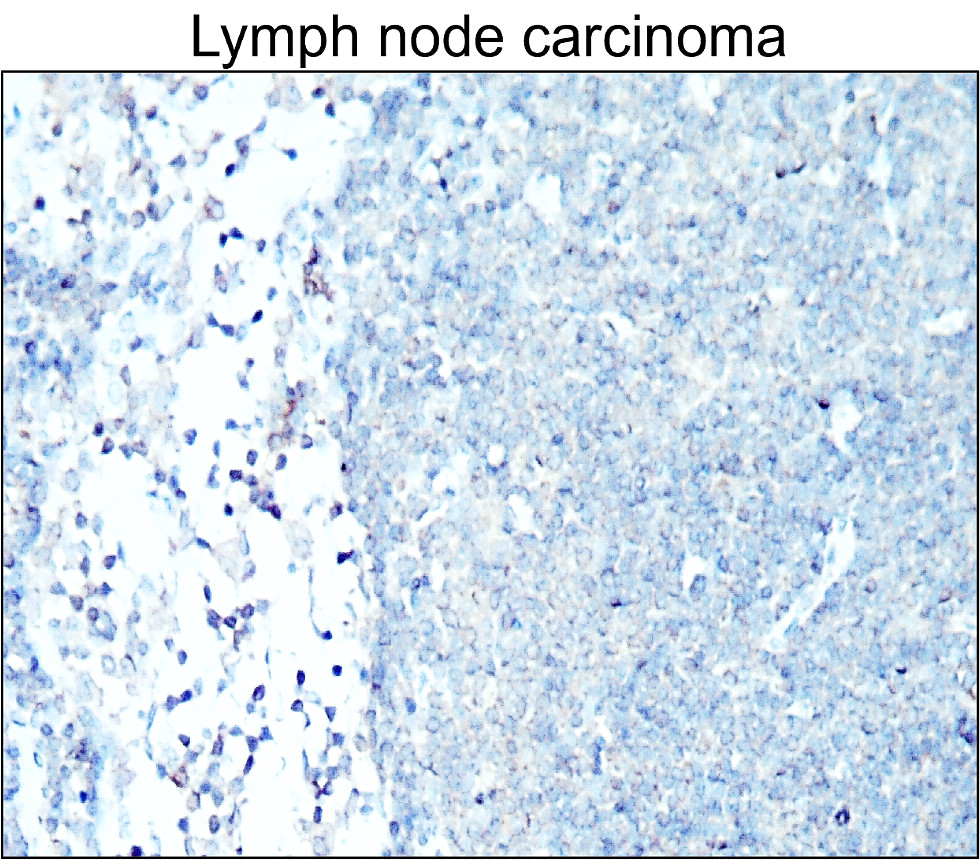
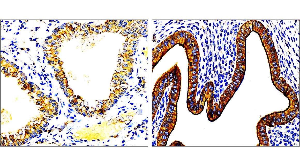
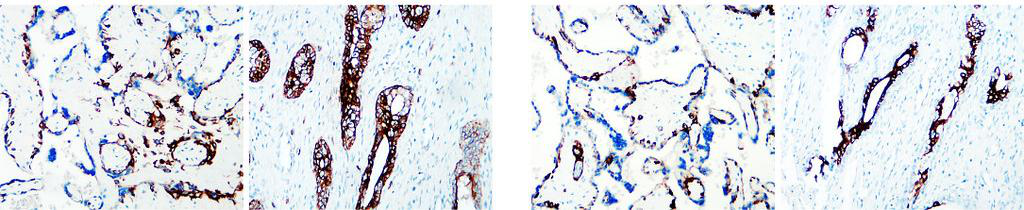
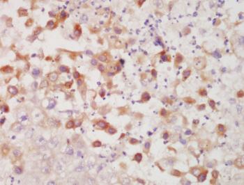
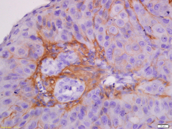
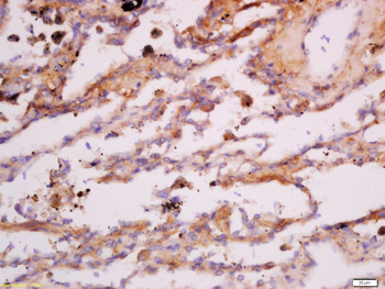
Tissue/Cell: Human esophageal carcinoma, 4% Paraformaldehyde-fixed and paraffin-embedded, Antigen retrieval: citrate buffer (0.01M, pH6.0), Boiling bathing for 15 min, Block endogenous peroxidase by 3% Hydrogen peroxide for 30 min, Blocking buffer (normal goat serum) at 37°C for 20 min, Incubation: Anti-CK7 Polyclonal Antibody, Unconjugated (orb10410) 1:200, overnight at 4°C, followed by conjugation to the secondary antibody and DAB staining.
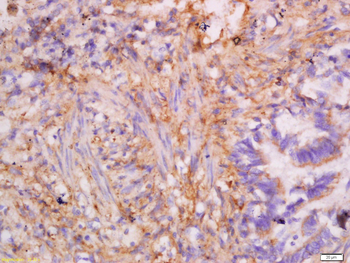
Tissue/Cell: human laryngocarcinoma, 4% Paraformaldehyde-fixed and paraffin-embedded, Antigen retrieval: citrate buffer (0.01M, pH6.0), Boiling bathing for 15 min, Block endogenous peroxidase by 3% Hydrogen peroxide for 30 min, Blocking buffer (normal goat serum) at 37°C for 20 min, Incubation: Anti-CK7 Polyclonal Antibody, Unconjugated (orb10410) 1:200, overnight at 4°C, followed by conjugation to the secondary antibody and DAB staining.
Tissue/Cell: human laryngocarcinoma, 4% Paraformaldehyde-fixed and paraffin-embedded, Antigen retrieval: citrate buffer (0.01M, pH6.0), Boiling bathing for 15 min, Block endogenous peroxidase by 3% Hydrogen peroxide for 30 min, Blocking buffer (normal goat serum) at 37°C for 20 min, Incubation: Anti-CK7 Polyclonal Antibody, Unconjugated (orb10410) 1:200, overnight at 4°C, followed by conjugation to the secondary antibody and DAB staining.
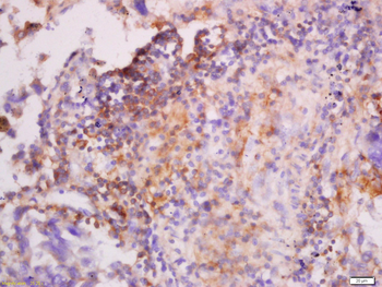
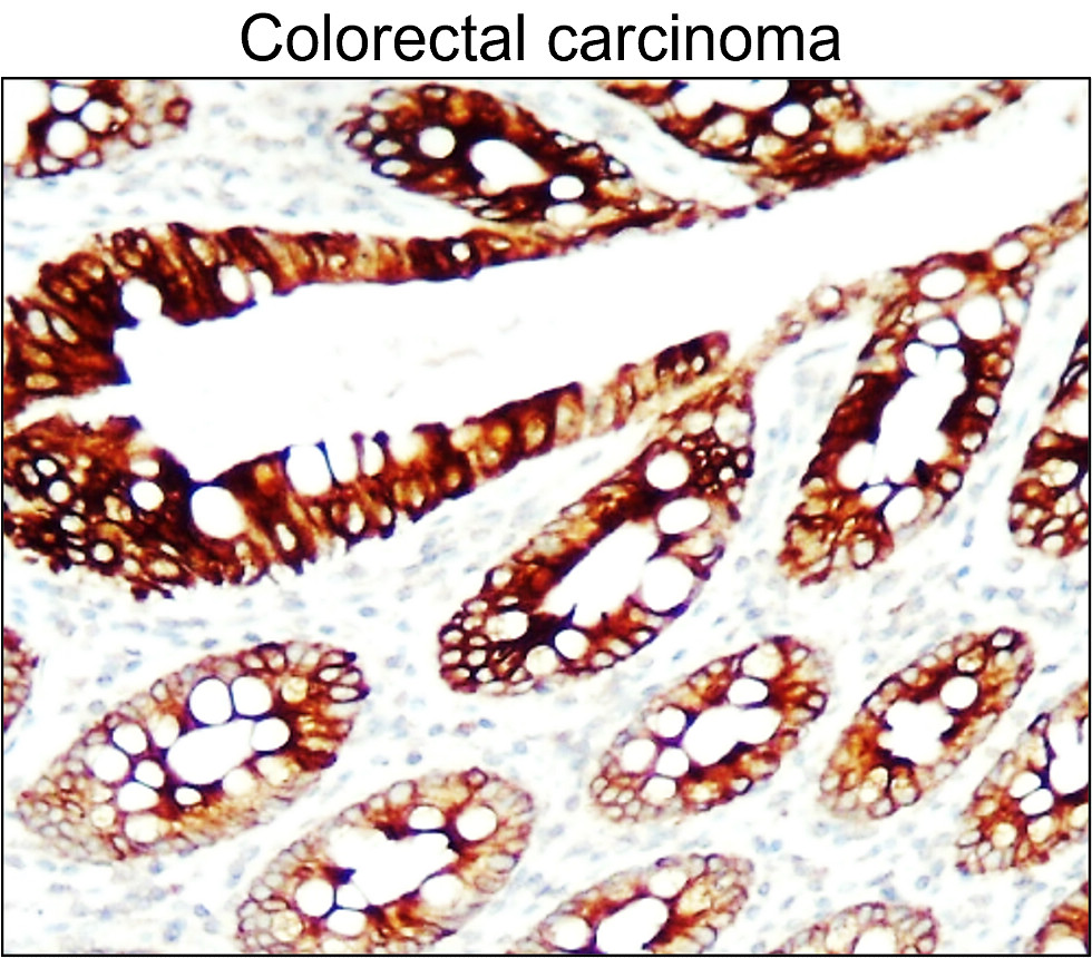
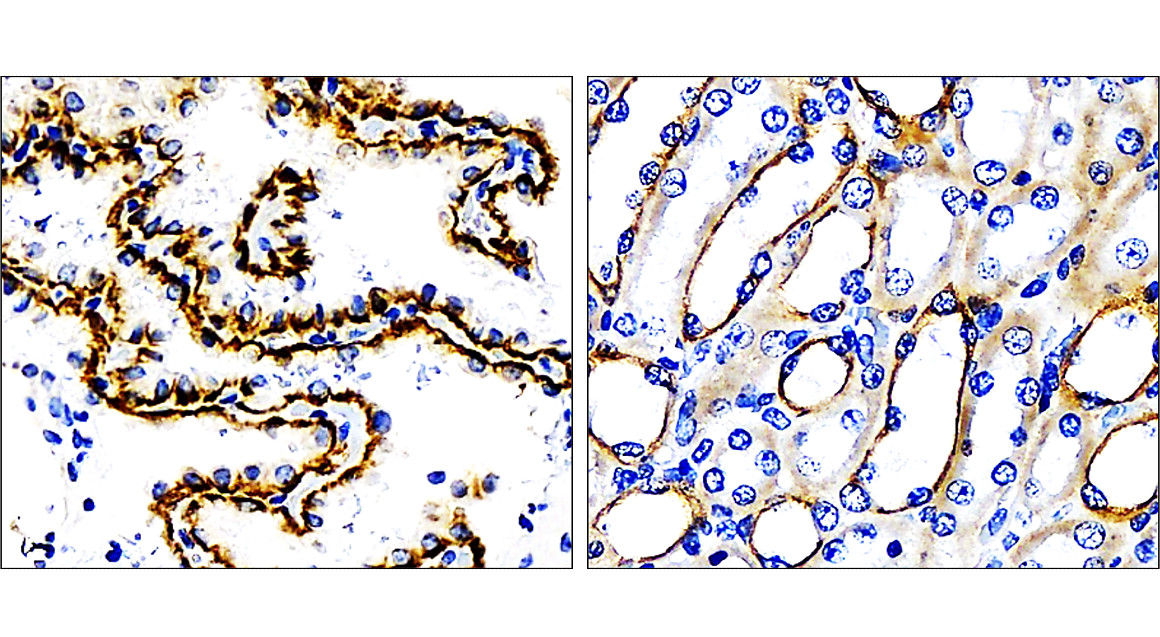
Tissue/Cell: human lung carcinoma, 4% Paraformaldehyde-fixed and paraffin-embedded, Antigen retrieval: citrate buffer (0.01M, pH6.0), Boiling bathing for 15 min, Block endogenous peroxidase by 3% Hydrogen peroxide for 30 min, Blocking buffer (normal goat serum) at 37°C for 20 min, Incubation: Anti-CK7 Polyclonal Antibody, Unconjugated (orb10410) 1:200, overnight at 4°C, followed by conjugation to the secondary antibody and DAB staining.
Tissue/Cell: human lung carcinoma, 4% Paraformaldehyde-fixed and paraffin-embedded, Antigen retrieval: citrate buffer (0.01M, pH6.0), Boiling bathing for 15 min, Block endogenous peroxidase by 3% Hydrogen peroxide for 30 min, Blocking buffer (normal goat serum) at 37°C for 20 min, Incubation: Anti-CK7 Polyclonal Antibody, Unconjugated (orb10410) 1:200, overnight at 4°C, followed by conjugation to the secondary antibody and DAB staining.
Tissue/Cell: human lung carcinoma, 4% Paraformaldehyde-fixed and paraffin-embedded, Antigen retrieval: citrate buffer (0.01M, pH6.0), Boiling bathing for 15 min, Block endogenous peroxidase by 3% Hydrogen peroxide for 30 min, Blocking buffer (normal goat serum) at 37°C for 20 min, Incubation: Anti-CK7 Polyclonal Antibody, Unconjugated (orb10410) 1:200, overnight at 4°C, followed by conjugation to the secondary antibody and DAB staining.
- Item 1 of 13
- Item 1 of 6
CK7 Rabbit Polyclonal Antibody [orb13342]
FC, ICC, IF, IHC-Fr, IHC-P, WB
Human, Rat
Human, Mouse, Rat
Rabbit
Polyclonal
Unconjugated
200 μl, 50 μl, 100 μl - Item 1 of 3
KRT7 Rabbit Polyclonal Antibody [orb583924]
IHC, WB
Bovine, Canine, Equine
Human
Rabbit
Polyclonal
Unconjugated
100 μl - Item 1 of 2
Keratin 7 Rabbit Polyclonal Antibody [orb77387]
ELISA, IHC, WB
Human, Mouse, Rat
Rabbit
Polyclonal
Unconjugated
100 μg - Item 1 of 2
Keratin 7 Rabbit Polyclonal Antibody [orb77388]
ELISA, IHC, WB
Rat
Human
Rabbit
Polyclonal
Unconjugated
100 μg