You have no items in your shopping cart.
Cart summary
Item 1 of 5
Item 1 of 5
CIRBP antibody
Catalog Number: orb20642

| Catalog Number | orb20642 |
|---|---|
| Category | Antibodies |
| Description | Goat polyclonal to CIRBP |
| Species/Host | Goat |
| Clonality | Polyclonal |
| Tested applications | ELISA, FC, IF, WB |
| Reactivity | Bovine, Canine, Human, Mouse, Rat |
| Dilution range | ELISA: 1:16000, WB: 0.05-0.2 μg/ml |
| Conjugation | Unconjugated |
| MW | 18.6 |
| Target | CIRBP (aa 81-91) |
| Entrez | 1153 |
| Protein Sequence | QAGKSSDNRSR |
| RRID | AB_10750386 |
| Storage | Aliquot and store at -20°C. Minimize freezing and thawing. |
| Buffer/Preservatives | Supplied at 0.5 mg/ml in Tris saline, 0.02% sodium azide, pH 7.3 with 0.5% bovine serum albumin. Aliquot and store at -20°C. Minimize freezing and thawing. |
| Alternative names | anti CIRBP antibody, anti cold inducible RNA bindi Read more... |
| Note | For research use only |
| Application notes | ELISA: Peptide ELISA: antibody detection limit dilution 1:16000.WB: Approx 18kDa band observed in Mouse and Rat Testis lysates (calculated MW of 18.6kDa according to Mouse NP_031731.1 and Rat NP_112409.2). Recommended concentration: 0.05-0.2 μg/ml. An additional band of unknown identity was also consistently observed at 23kDa. This band was successfully blocked by incubation with the immunizing peptide. We would appreciate any feedback from people in the field - have any such results been reported with other antibodies/lysates Have any further splice variants/modified forms been reported |
| Expiration Date | 12 months from date of receipt. |

orb20642 (0.05ug/ml) staining of Mouse Testis lysate (35ug protein in RIPA buffer). Primary incubation was 1 hour. Detected by chemiluminescence.
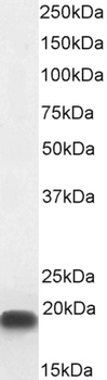
orb20642 (0.03 µg/mL) staining of MCF7 nuclear cell lysate (35 µg protein in RIPA buffer). Detected by chemiluminescence.
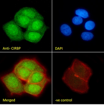
orb20642 Immunofluorescence analysis of paraformaldehyde fixed MCF7 cells, permeabilized with 0.15% Triton. Primary incubation 1 hr (10 µg/mL) followed by Alexa Fluor 488 secondary antibody (2 µg/mL), showing nuclear staining. Actin filaments were stained with phalloidin (red) and the nuclear stain is DAPI (blue). Negative control: Unimmunized goat IgG (10 µg/mL) followed by Alexa Fluor 488 secondary antibody (2 µg/mL).
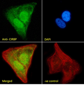
orb20642 Immunofluorescence analysis of paraformaldehyde fixed U2OS cells, permeabilized with 0.15% Triton. Primary incubation 1 hr (10 µg/mL) followed by Alexa Fluor 488 secondary antibody (2 µg/mL), showing nuclear staining. Actin filaments were stained with phalloidin (red) and the nuclear stain is DAPI (blue). Negative control: Unimmunized goat IgG (10 µg/mL) followed by Alexa Fluor 488 secondary antibody (2 µg/mL).
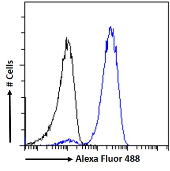
orb20642 Flow cytometric analysis of paraformaldehyde fixed MCF7 cells (blue line), permeabilized with 0.5% Triton. Primary incubation 1 hr (10 µg/mL) followed by Alexa Fluor 488 secondary antibody (1 µg/mL). IgG control: Unimmunized goat IgG (black line) followed by Alexa Fluor 488 secondary antibody.
- Item 1 of 7
- Item 1 of 3
CIRBP antibody [orb377997]
IF, IH, WB
Human, Mouse, Rat
Rabbit
Polyclonal
Unconjugated
100 μl, 200 μl, 50 μl - Item 1 of 3
- Item 1 of 3
- Item 1 of 3
CIRBP antibody [orb247495]
ICC, IF, IHC, WB
Human, Mouse, Rat
Polyclonal
Unconjugated
200 μl, 100 μl, 50 μl


Submit a review
Filter by Rating

- 5 stars

- 4 stars
- 3 stars
- 2 stars
- 1 stars
