You have no items in your shopping cart.
Cart summary
Item 1 of 2
Item 1 of 2
CENP-Q antibody
Catalog Number: orb344649

| Catalog Number | orb344649 |
|---|---|
| Category | Antibodies |
| Description | CENP-Q antibody |
| Species/Host | Rabbit |
| Clonality | Polyclonal |
| Tested applications | ELISA, IF, Multiplex Assay, WB |
| Reactivity | Human |
| Isotype | IgG |
| Immunogen | This protein A purified antibody was prepared from whole rabbit serum produced by repeated immunizations with full-length human CENP-Q recombinant protein. |
| Concentration | 1.15 mg/mL |
| Dilution range | ELISA: 1:5,000 1:20,000, IF: User Optimized, WB: 1:100 - 1:500 |
| Form/Appearance | Liquid (sterile filtered) |
| Purity | This product was protein A purified from monospecific antiserum by immunoaffinity chromatography using protein A coupled to agarose beads. This antibody is specific for human CENP-Q protein. A BLAST analysis of the full length sequence was used to suggest partial cross-reactivity with CENP-Q based on the following percentage homologies: macaque (91%), horse (76%), bovine (74%), dog (73%), swine (71%), mouse (65%) and rat (60%). Cross-reactivity with CENP-Q from other sources has not been determined. |
| Conjugation | Unconjugated |
| UniProt ID | Q7L2Z9 |
| NCBI | 40068061 |
| Storage | Store vial at -20° C or below prior to opening. This vial contains a relatively low volume of reagent (25 µL). To minimize loss of volume dilute 1:10 by adding 225 µL of the buffer stated above directly to the vial. Recap, mix thoroughly and briefly centrifuge to collect the volume at the bottom of the vial. Use this intermediate dilution when calculating final dilutions as recommended below. Store the vial at -20°C or below after dilution. Avoid cycles of freezing and thawing. |
| Buffer/Preservatives | 0.01% (w/v) Sodium Azide |
| Alternative names | rabbit anti-CENP-Q Antibody, CenpQ, centromere pro Read more... |
| Note | For research use only |
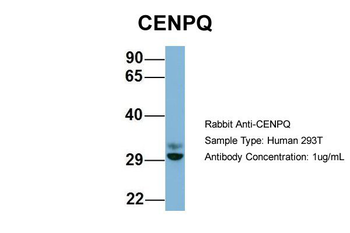
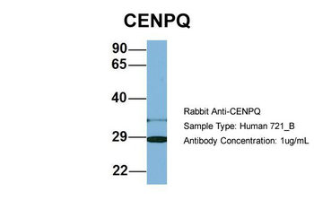
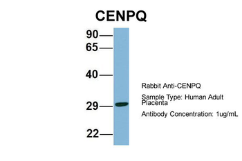
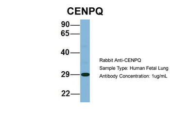
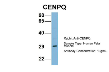
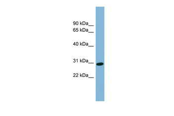
| Application notes | This protein A purified antibody has been tested for use in ELISA, immunofluorescence microscopy and western blotting. Specific conditions for reactivity should be optimized by the end user. Expect a band approximately 26-31 kDa in size corresponding to human CENP-Q by western blotting in the appropriate cell lysate or extract. |
| Expiration Date | 12 months from date of receipt. |
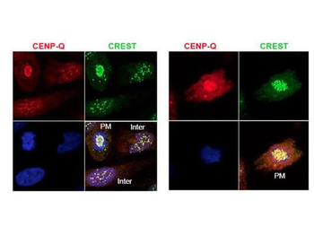
Immunofluorescence microscopy using Biorbyt's protein A purified anti-CENP-Q antibody shows detection of endogenous CENP-Q in HeLa whole cell lysate. Primary antibody was used at 1:100 followed by secondary antibody diluted 1:150. Red punctate anti-CENP-Q signal colocalizes in overlay images with green punctate anti-CREST signals at the kinetochores (attached points of sister chromatids). Visible are colocalized CENP-Q and CREST signal at various stages of the cell cycle as indicated from interphase to the end of mitosis. Nuclei are counter stained with bisbenzimide.
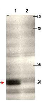
Western blot using Biorbyt's protein A purified anti-CENP-Q antibody shows detection of endogenous CENP-Q in a HeLa whole cell lysate (lane 1, arrowhead). The blot was incubated for 1.5 hours at room temperature using the primary antibody diluted to 0.5 µg/mL, followed by washes and incubation with to the secondary antibody. Lane 1: Lysates from HeLa cells transfected with control sh-virus wherein the expression of CENP-Q is expected to not to alter. Lane 2: Lysates from HeLa cells transfected with Cenp-Q sh-virus wherein the expression of CENP-Q is knocked down significantly to a level where it is not being detected at all under the tested condition/WB exposure time.
- Item 1 of 6
- Item 1 of 2
- Item 1 of 2
- Item 1 of 2
- Item 1 of 2


Submit a review
Filter by Rating

- 5 stars

- 4 stars
- 3 stars
- 2 stars
- 1 stars
