You have no items in your shopping cart.
Cart summary
Item 1 of 2
Item 1 of 2
CENP-Q antibody
Catalog Number: orb344648

| Catalog Number | orb344648 |
|---|---|
| Category | Antibodies |
| Description | CENP-Q antibody |
| Species/Host | Rabbit |
| Clonality | Polyclonal |
| Tested applications | ELISA, IF, Multiplex Assay, WB |
| Reactivity | Human |
| Isotype | IgG |
| Immunogen | This protein A purified antibody was prepared from whole rabbit serum produced by repeated immunizations with full-length human CENP-Q recombinant protein. |
| Concentration | 1.15 mg/mL |
| Dilution range | ELISA: 1:5,000 1:20,000, IF: User Optimized, WB: 1:100 - 1:500 |
| Form/Appearance | Liquid (sterile filtered) |
| Purity | This product was protein A purified from monospecific antiserum by immunoaffinity chromatography using protein A coupled to agarose beads. This antibody is specific for human CENP-Q protein. A BLAST analysis of the full length sequence was used to suggest partial cross-reactivity with CENP-Q based on the following percentage homologies: macaque (91%), horse (76%), bovine (74%), dog (73%), swine (71%), mouse (65%) and rat (60%). Cross-reactivity with CENP-Q from other sources has not been determined. |
| Conjugation | Unconjugated |
| UniProt ID | Q7L2Z9 |
| NCBI | 40068061 |
| Storage | Store vial at -20° C prior to opening. Aliquot contents and freeze at -20° C or below for extended storage. Avoid cycles of freezing and thawing. Centrifuge product if not completely clear after standing at room temperature. This product is stable for several weeks at 4° C as an undiluted liquid. Dilute only prior to immediate use. |
| Buffer/Preservatives | 0.01% (w/v) Sodium Azide |
| Alternative names | rabbit anti-CENP-Q Antibody, CenpQ, centromere pro Read more... |
| Note | For research use only |
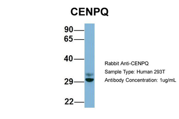
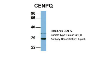
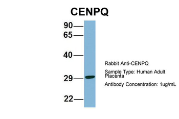
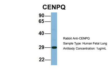
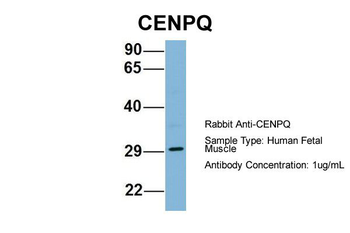
| Application notes | This protein A purified antibody has been tested for use in ELISA, immunofluorescence microscopy and western blotting. Specific conditions for reactivity should be optimized by the end user. Expect a band approximately 26-31 kDa in size corresponding to human CENP-Q by western blotting in the appropriate cell lysate or extract. |
| Expiration Date | 12 months from date of receipt. |

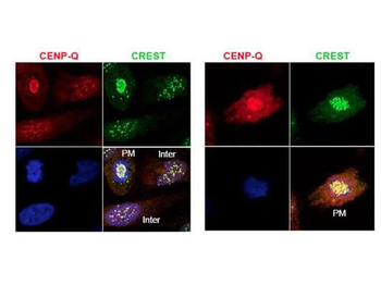
Immunofluorescence microscopy using Biorbyt's protein A purified anti-CENP-Q antibody shows detection of endogenous CENP-Q in HeLa whole cell lysate. Primary antibody was used at 1:100 followed by secondary antibody diluted 1:150. Red punctate anti-CENP-Q signal colocalizes in overlay images with green punctate anti-CREST signals at the kinetochores (attached points of sister chromatids). Visible are colocalized CENP-Q and CREST signal at various stages of the cell cycle as indicated from interphase to the end of mitosis. Nuclei are counter stained with bisbenzimide.

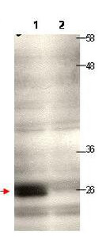
Western blot using Biorbyt's protein A purified anti-CENP-Q antibody shows detection of endogenous CENP-Q in a HeLa whole cell lysate (lane 1, arrowhead). The blot was incubated for 1.5 hours at room temperature using the primary antibody diluted to 0.5 µg/mL, followed by washes and incubation with to the secondary antibody. Lane 1: Lysates from HeLa cells transfected with control sh-virus wherein the expression of CENP-Q is expected to not to alter. Lane 2: Lysates from HeLa cells transfected with Cenp-Q sh-virus wherein the expression of CENP-Q is knocked down significantly to a level where it is not being detected at all under the tested condition/WB exposure time.
- Item 1 of 6
- Item 1 of 2
- Item 1 of 2
- Item 1 of 2
- Item 1 of 2


Submit a review
Filter by Rating

- 5 stars

- 4 stars
- 3 stars
- 2 stars
- 1 stars
