You have no items in your shopping cart.
Cart summary
Item 1 of 2
Item 1 of 2
CENP-A Antibody: FITC
Catalog Number: orb148111

| Catalog Number | orb148111 |
|---|---|
| Category | Antibodies |
| Description | Mouse monoclonal to CENP-A (FITC). A replicated chromosome includes two kinetochores that control chromosome segregation during mitosis. The Centromere Protein-A, CENP-A, is a Histone H3-like protein that contains a C-terminal H3-like domain, which is required for centromere localization of CENP-A, and an antigenic N-terminal domain. CENP-A, originally isolated from HeLa cells, is essential for kinetochore targeting of CENP-C. In the presence of DNA CENP-A forms an octa-meric complex with histones H4, H2A, H2B. CENP-A specifically localizes to active centromeres and is a component of specialized centromeric nucleosomes, on which kinetochores are assembled. CENP-A is essential for nucleosomal packaging of centromeric DNA at interphase and functions as a centromere formation marker on the chromosome.. |
| Species/Host | Mouse |
| Clonality | Monoclonal |
| Clone Number | 5A7-2E11 |
| Tested applications | ICC, IF, IHC |
| Reactivity | Human |
| Isotype | IgG1 |
| Immunogen | Synthetic peptide corresponding to a portion of human CENP-A |
| Concentration | 1 mg/ml |
| Dilution range | WB (1:1000), ICC/IF (1:100) |
| Conjugation | FITC |
| MW | 18kDa |
| Target | CENPA |
| Entrez | 1058 |
| UniProt ID | P49450 |
| NCBI | NP_001035891.1 |
| Storage | Conjugated antibodies should be stored according to the product label |
| Buffer/Preservatives | 640.91mM DMSO, 136.36mM Ethanolamine, 9.09mM Sodium Bicarbonate in 90.9% PBS |
| Alternative names | CENP A antibody, cenpa antibody, Centromere auto a Read more... |
| Note | For research use only |
| Application notes | 1 µg/ml of SMC-202 was sufficient for detection of CENPA in 20 µg of U2OS cell lysate by colorimetric immunoblot analysis using Goat anti-mouse IgG:HRP as the secondary antibody. |
| Expiration Date | 12 months from date of receipt. |

Immunocytochemistry/Immunofluorescence analysis using Mouse Anti-CENP-A Monoclonal Antibody, Clone 5A7-2E11. Tissue: Colon cancer cell line (HT-29). Species: Human. Fixation: 4% Formaldehyde for 15 min at RT. Primary Antibody: Mouse Anti-CENP-A Monoclonal Antibody at 1:100 for 60 min at RT. Secondary Antibody: Goat Anti-Mouse ATTO 488 at 1:100 for 60 min at RT. Counterstain: DAPI (blue) nuclear stain at 1:5000 for 5 min RT. Localization: Nucleus. Magnification: 60X.
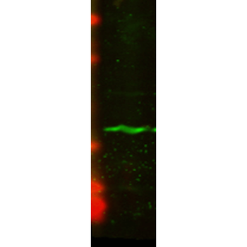
Western Blot analysis of Human U2OS cell lysate showing detection of CENP-A protein using Mouse Anti-CENP-A Monoclonal Antibody, Clone 5A7-2E11. Primary Antibody: Mouse Anti-CENP-A Monoclonal Antibody at 1:1000.

Centromeric histone H3-like protein-2 Rabbit Polyclonal Antibody (FITC) [orb15760]
IF
Plant
Rabbit
Polyclonal
FITC
100 μlCentromeric histone H3-like protein-2 Rabbit Polyclonal Antibody (FITC) [orb15762]
IF
Plant
Rabbit
Polyclonal
FITC
100 μlCENPA Rabbit Polyclonal Antibody (FITC) [orb2117349]
WB
Bovine, Canine, Equine, Goat, Guinea pig, Human, Mouse, Rabbit, Rat, Zebrafish
Rabbit
Polyclonal
FITC
100 μlCENPA Rabbit Polyclonal Antibody (FITC) [orb2117352]
IHC, WB
Human, Mouse, Rat
Rabbit
Polyclonal
FITC
100 μl
