You have no items in your shopping cart.
Cart summary
Item 1 of 5
Item 1 of 5
CDH4 Antibody (N-term)
Catalog Number: orb1936212

| Catalog Number | orb1936212 |
|---|---|
| Category | Antibodies |
| Description | Affinity Purified Rabbit Polyclonal Antibody (Pab) |
| Species/Host | Rabbit |
| Clonality | Polyclonal |
| Clone Number | RB13654 |
| Tested applications | FC, IF, IHC-P, WB |
| Predicted Reactivity | Mouse, Rat |
| Reactivity | Human |
| Isotype | Rabbit IgG |
| Antibody Type | Primary Antibody |
| Dilution range | IF: 1:10~50, WB: 1:2000, WB: 1:2000, IHC-P: 1:10~50, FC: 1:10~50 |
| Form/Appearance | Purified polyclonal antibody supplied in PBS with 0.09% (W/V) sodium azide. This antibody is purified through a protein A column, followed by peptide affinity purification. |
| Conjugation | Unconjugated |
| MW | 100281 Da |
| Target | This CDH4 antibody is generated from rabbits immunized with a KLH conjugated synthetic peptide between 175-203 amino acids from the N-terminal region of human CDH4. |
| UniProt ID | P55283 |
| NCBI | NP_001785.2, NP_001239267.1, NP_001239268.1 |
| Storage | Maintain refrigerated at 2-8°C for up to 2 weeks. For long term storage store at -20°C in small aliquots to prevent freeze-thaw cycles |
| Alternative names | Cadherin-4, Retinal cadherin, R-CAD, R-cadherin, C Read more... |
| Note | For research use only |
| Expiration Date | 12 months from date of receipt. |

Flow cytometric analysis of HepG2 cells using CDH4 Antibody (N-term) (bottom histogram) compared to a negative control cell (top histogram). FITC-conjugated goat-anti-rabbit secondary antibodies were used for the analysis.
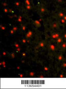
Immunofluorescence analysis of CDH4 Antibody (N-term) Antibody with paraffin-embedded human brain tissue.0.025 mg/ml primary antibody was followed by FITC-conjugated goat anti-rabbit lgG (whole molecule). FITC emits green fluorescence.Red counterstaining is PI.
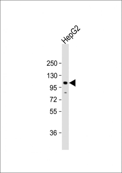
Anti-CDH4 Antibody (N-term) at 1:2000 dilution + HepG2 whole cell lysate. Lysates/proteins at 20 µg per lane. Secondary Goat Anti-Rabbit IgG, (H+L), Peroxidase conjugated at 1/10000 dilution. Predicted band size: 100 kDa. Blocking/Dilution buffer: 5% NFDM/TBST.
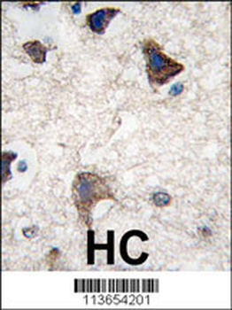
Formalin-fixed and paraffin-embedded human brain tissue reacted with CDH4 antibody (N-term), which was peroxidase-conjugated to the secondary antibody, followed by DAB staining. This data demonstrates the use of this antibody for immunohistochemistry; clinical relevance has not been evaluated.
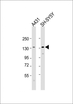
All lanes: Anti-CDH4 Antibody (N-term) at 1:2000 dilution. Lane 1: A431 whole cell lysate. Lane 2: SH-SY5Y whole cell lysate. Lysates/proteins at 20 µg per lane. Secondary Goat Anti-Rabbit IgG, (H+L), Peroxidase conjugated at 1/10000 dilution. Predicted band size: 100 kDa. Blocking/Dilution buffer: 5% NFDM/TBST.
