You have no items in your shopping cart.
Cart summary
Item 1 of 4
Item 1 of 4
CD74 Antibody: PerCP
Catalog Number: orb146943

| Catalog Number | orb146943 |
|---|---|
| Category | Antibodies |
| Description | Mouse monoclonal to CD74 (PerCP). CD74 is a non-polymorphic type II integral membrane protein. It has a short N-terminal cytoplasmic tail of 28 amino acids, followed by a single 24-aa transmembrane region and an approximately 150-aa lumenal domain. The CD74 chain is thought to function mainly as an MHC class II chaperone, which promotes ER exit of MHC class II molecules, directs them to endocytic compartments, prevents peptide binding in the ER, and contributes to peptide editing in the MHC class II compartment. Class II MHC and Ii expression was believed to be restricted to classical antigen-presenting cells (APC); however, during inflammation, other cell types, including mucosal epithelial cells, have also been reported to express class II MHC molecules. Experiments that investigate cell-surface CD74 are complicated by the fact that CD74 remains on the cell surface for a very short time. The surface half-life of CD74 was calculated to be fewer than 10 minutes. CD74 however has also recently been shown to have a role as an accessory-signaling molecule because of its high-affinity binding to the pro-inflammatory cytokine, macrophage migration-inhibitory factor (MIF). The restricted expression of CD74 by normal tissues and its very rapid internalization make CD74 an attractive therapeutic target for both cancer and immunologic diseases.. |
| Species/Host | Mouse |
| Clonality | Monoclonal |
| Clone Number | PIN.1 |
| Tested applications | ELISA, ICC, IF, IHC, WB |
| Reactivity | Human, Mouse |
| Isotype | IgG1 |
| Immunogen | Human CD74 invariant chain synthetic peptide |
| Concentration | 1 mg/ml |
| Dilution range | WB (1:1000), IHC (1:100), ICC/IF (1:50) |
| Conjugation | PerCP |
| MW | 33-35kDa |
| Target | CD74 |
| Entrez | 972 |
| UniProt ID | P04233 |
| NCBI | NP_001020329.1 |
| Storage | Conjugated antibodies should be stored according to the product label |
| Buffer/Preservatives | 95.64mM Phosphate, 2.48mM MES and 2mM EDTA |
| Alternative names | DHLAG antibody, HLA DR gamma antibody, HLADG antib Read more... |
| Note | For research use only |
| Application notes | 1 µg/ml of SMC-116 was sufficient for detection of CD74 in 20 µg of PALA cell lysates by colorimetric immunolot analysis using goat anti-mouse IgG: AP as the secondary antibody. |
| Expiration Date | 12 months from date of receipt. |

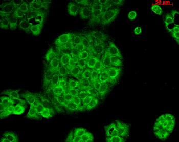
Immunocytochemistry/Immunofluorescence analysis using Mouse Anti-CD74 Monoclonal Antibody, Clone PIN 1.1. Tissue: Cervical cancer cell line (HeLa). Species: Human. Fixation: 2% Formaldehyde for 20 min at RT. Primary Antibody: Mouse Anti-CD74 Monoclonal Antibody at 1:100 for 12 hours at 4°C. Secondary Antibody: FITC Goat Anti-Mouse (green) at 1:200 for 2 hours at RT. Counterstain: DAPI (blue) nuclear stain at 1:40000 for 2 hours at RT. Localization: Cell membrane. Endoplasmic reticulum membrane. Golgi apparatus. Endosome. Lysosome. Magnification: 100x. (A) DAPI (blue) nuclear stain. (B) Anti-CD74 Antibody. (C) Composite.
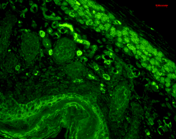
Immunohistochemistry analysis using Mouse Anti-CD74 Monoclonal Antibody, Clone PIN 1.1. Tissue: backskin. Species: Mouse. Fixation: Bouin's Fixative and paraffin-embedded. Primary Antibody: Mouse Anti-CD74 Monoclonal Antibody at 1:100 for 1 hour at RT. Secondary Antibody: FITC Goat Anti-Mouse (green) at 1:50 for 1 hour at RT. Localization: Beautiful basal to suprabasal staining in epidermis, dermis, hair follicles and muscle.
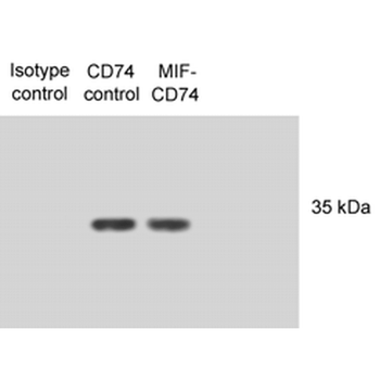
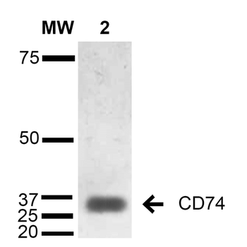
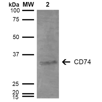
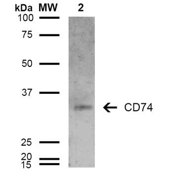
Western Blot analysis of Human N87 cell lysates showing detection of CD74 protein using Mouse Anti-CD74 Monoclonal Antibody, Clone PIN 1.1. Primary Antibody: Mouse Anti-CD74 Monoclonal Antibody at 1:1000. Lysates treated with macrophage inhibitory factor (MIF).
Immunocytochemistry/Immunofluorescence analysis using Mouse Anti-CD74 Monoclonal Antibody, Clone PIN 1.1. Tissue: HaCaT cells. Species: Human. Fixation: Cold 100% methanol for 10 minutes at -20°C. Primary Antibody: Mouse Anti-CD74 Monoclonal Antibody at 1:100 for 1 hour at RT. Secondary Antibody: FITC Goat Anti-Mouse (green) at 1:50 for 1 hour at RT. Localization: Cytoplasmic Staining.
- Item 1 of 2
- Item 1 of 2
- Item 1 of 2

CD74 Rabbit Polyclonal Antibody (PerCP-Cy5.5) [orb2438290]
IF
Mouse, Rat
Mouse
Rabbit
Polyclonal
PerCP/Cy5.5
100 μl