You have no items in your shopping cart.
Cart summary
Item 1 of 2
Item 1 of 2
CD59 Antibody
Catalog Number: orb749914

| Catalog Number | orb749914 |
|---|---|
| Category | Antibodies |
| Description | Reacts with human CD59, a 20kDa glycosyl phosphatidyl-inositol (GPI)-anchored cell surface protein. CD59 regulates complement-mediated cell lysis, and it is involved in lymphocyte signal transduction. This protein is a potent inhibitor of the complement membrane attack complex, whereby it binds complement C8 and/or C9 during the assembly of this complex, thereby inhibiting the incorporation of multiple copies of C9 into the complex, which is necessary for osmolytic pore formation. It inhibits formation of MAC, thus protecting cells from complement-mediated lysis. Genetic defects in GPI-anchor attachment, that cause a reduction or loss of CD59 and CD55 on erythrocytes produce the symptoms of the disease paroxysmal hemoglobinuria (PNH). This MAb is useful for study on GPI-anchored proteins, PNH and CD59 functions. CD59 is widely distributed on cells in all tissues. The expression of CD59 on erythrocytes is important for their survival. |
| Species/Host | Mouse |
| Clonality | Monoclonal |
| Clone Number | MACIF/629 |
| Tested applications | IHC-P |
| Reactivity | Human |
| Isotype | Mouse IgG1, kappa |
| Immunogen | Recombinant full-length human protein was used as the immunogen for the CD59 antibody. |
| Dilution range | Immunohistochemistry (FFPE): 1-2ug/ml for 30 min at RT |
| Purity | Protein G affinity chromatography |
| Conjugation | Unconjugated |
| Formula | 0.2 mg/ml in 1X PBS with 0.1 mg/ml BSA (US sourced) and 0.05% sodium azide |
| Hazard Information | This CD59 antibody is available for research use only. |
| UniProt ID | P13987 |
| Storage | Store the CD59 antibody at 2-8°C (with azide) or aliquot and store at -20°C or colder (without azide). |
| Buffer/Preservatives | 0.2 mg/ml in 1X PBS with 0.1 mg/ml rAlbumin (US sourced) and 0.05% sodium azide |
| Note | For research use only |
| Application notes | Optimal dilution of the CD59 antibody should be determined by the researcher.1. Staining of formalin-fixed tissues is enhanced by boiling tissue sections in 10mM Citrate buffer, pH 6.0, for 10-20 min followed by cooling at RT for 20 min.2. The prediluted format is supplied in a dropper bottle and is optimized for use in IHC. After epitope retrieval step (if required), drip mAb solution onto the tissue section and incubate at RT for 30 min. |
| Expiration Date | 12 months from date of receipt. |
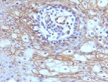
Formalin-fixed, paraffin-embedded human tongue stained with CD59 antibody (MACIF/629)
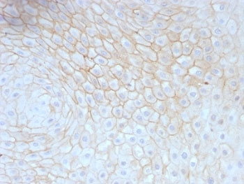
Formalin-fixed, paraffin-embedded human tonsil stained with CD59 antibody (MACIF/629)
- Item 1 of 6
- Item 1 of 5
CD59 Antibody(monoclonal, 3C10) [orb623829]
FC, ICC, IF, IHC, WB
Human
Mouse
Monoclonal
Unconjugated
10 μg, 100 μg - Item 1 of 4
- Item 1 of 4
- Item 1 of 4
CD59 glycoprotein CD59 Antibody [orb308756]
FC, ICC, IF, IHC, WB
Human
Rabbit
Polyclonal
Unconjugated
10 μg, 100 μg


Submit a review
Filter by Rating

- 5 stars

- 4 stars
- 3 stars
- 2 stars
- 1 stars
