You have no items in your shopping cart.
Cart summary
Item 1 of 5
Item 1 of 5
CD56 Rabbit Polyclonal Antibody
Catalog Number: orb10339

| Catalog Number | orb10339 |
|---|---|
| Category | Antibodies |
| Description | CD56 Rabbit Polyclonal Antibody |
| Species/Host | Rabbit |
| Clonality | Polyclonal |
| Tested applications | FC, ICC, IF, IHC-Fr, IHC-P, WB |
| Predicted Reactivity | Bovine, Canine, Equine, Gallus, Guinea pig, Porcine, Rabbit |
| Reactivity | Human, Mouse, Rat |
| Isotype | IgG |
| Immunogen | KLH conjugated synthetic peptide derived from human CD56 (621-720/858aa) |
| Antibody Type | Primary Antibody |
| Concentration | 1mg/ml |
| Dilution range | WB=1:500-2000, IHC-P=1:100-500, IHC-F=1:100-500, ICC/IF=1:100, IF=1:100-500, Flow-Cyt=1μg/Test |
| Form/Appearance | Liquid |
| Conjugation | Unconjugated |
| MW | 92 kDa |
| Target | NCAM1 |
| UniProt ID | P13591 |
| RRID | AB_10939981 |
| Storage | Maintain refrigerated at 2-8°C for up to 2 weeks. For long term storage store at -20°C in small aliquots to prevent freeze-thaw cycles. |
| Buffer/Preservatives | 0.01M TBS (pH7.4) with 1% rAlbumin, 0.02% Proclin300 and 50% Glycerol. |
| Alternative names | CD56 antigen; Cell Adhesion Molecule Neural 1; MSK Read more... |
| Note | For research use only |
| Expiration Date | 12 months from date of receipt. |

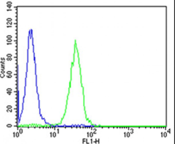
Blank control: Jurkat cells (blue). Primary Antibody: Rabbit Anti-CD56 antibody (orb10339), Dilution: 1 µg in 100 µl 1X PBS containing 0.5% BSA, Isotype Control Antibody: Rabbit IgG (orange), used under the same conditions, Secondary Antibody: Goat anti-rabbit IgG-PE (white blue), Dilution: 1:200 in 1 X PBS containing 0.5% BSA. Protocol, The cells were fixed with 2% paraformaldehyde (10 min). Primary antibody (orb10339, 1 µg/1x10^6 cells) were incubated for 30 min on the ice, followed by 1 X PBS containing 0.5% BSA + 1 0% goat serum (15 min) to block non-specific protein-protein interactions. Then the Goat Anti-rabbit IgG/PE antibody was added into the blocking buffer mentioned above to react with the primary antibody at 1/200 dilution for 30 min on ice. Acquisition of 20000 events was performed.
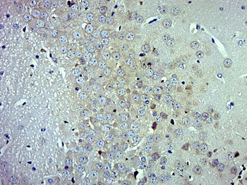
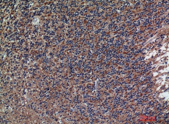
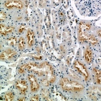
Paraformaldehyde-fixed, paraffin embedded (Mouse brain), Antigen retrieval by boiling in sodium citrate buffer (pH6.0) for 15 min, Block endogenous peroxidase by 3% hydrogen peroxide for 20 minutes, Blocking buffer (normal goat serum) at 37°C for 30 min, Antibody incubation with (CD56) Polyclonal Antibody, Unconjugated (orb10339) at 1:500 overnight at 4°C, followed by a conjugated secondary for 20 minutes and DAB staining.

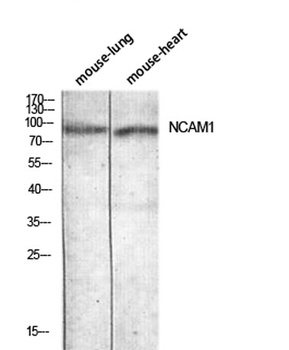
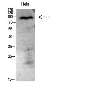
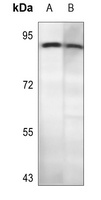
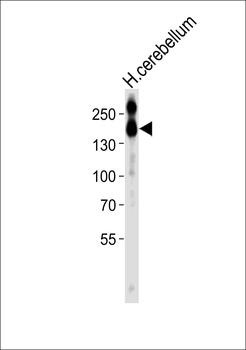
Sample: Cerebral cortex (Mouse) Lysate at 40 ug, Primary: Anti-CD56 (orb10339) at 1/1000 dilution, Secondary: IRDye800CW Goat Anti-Rabbit IgG at 1/20000 dilution, Predicted band size: 92 kD, Observed band size: 95 kD.
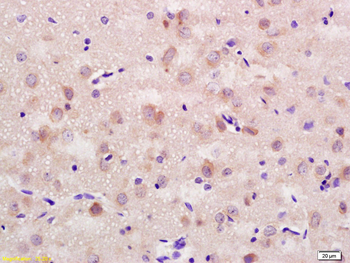
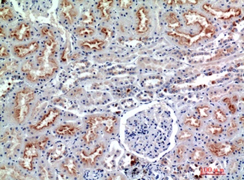
Tissue/Cell: rat brain tissue, 4% Paraformaldehyde-fixed and paraffin-embedded, Antigen retrieval: citrate buffer (0.01M, pH6.0), Boiling bathing for 15 min, Block endogenous peroxidase by 3% Hydrogen peroxide for 30 min, Blocking buffer (normal goat serum) at 37°C for 20 min, Incubation: Anti-CD56/NCAM1 Polyclonal Antibody, Unconjugated (orb10339) 1:200, overnight at 4°C, followed by conjugation to the secondary antibody and DAB staining.
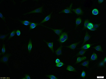
Tissue/Cell: SH-SY5Y cell, 4% Paraformaldehyde-fixed, Triton X-100 at room temperature for 20 min, Blocking buffer (normal goat serum) at 37°C for 20 min, Antibody incubation with (CD56) polyclonal Antibody, Unconjugated (orb10339) 1:100, 90 minutes at 37°C, followed by a FITC conjugated Goat Anti-Rabbit IgG antibody at 37°C for 90 minutes, DAPI (blue) was used to stain the cell nuclei.
- Item 1 of 4

CD56 Rabbit Polyclonal Antibody (PE) [orb485686]
FC, ICC, IF
Bovine, Canine, Equine, Gallus, Guinea pig, Porcine, Rabbit
Human, Mouse, Rat
Rabbit
Polyclonal
PE
100 μl- Item 1 of 3
NCAM1 Antibody (C-term) [orb1788087]
FC, IHC-P, WB
Mouse
Human, Rat
Rabbit
Polyclonal
Unconjugated
100 μl - Item 1 of 2
Anti-CD56 Antibody [orb315805]
IH, WB
Human, Mouse, Primate, Rat
Rabbit
Polyclonal
Unconjugated
200 μl, 100 μl, 30 μl - Item 1 of 3