You have no items in your shopping cart.
Cart summary
Item 1 of 8
Item 1 of 8
CD32b antibody
Catalog Number: orb19010

| Catalog Number | orb19010 |
|---|---|
| Category | Antibodies |
| Description | Goat polyclonal antibody to CD32b |
| Species/Host | Goat |
| Clonality | Polyclonal |
| Tested applications | ELISA, IF, IHC, WB |
| Reactivity | Human |
| Dilution range | ELISA: 1:32000, WB: 0.3-1 μg/ml |
| Conjugation | Unconjugated |
| MW | 34.0; 31.9; 31.9; 34.0; 33.4 |
| Target | CD32 / FCGR2B |
| Entrez | 2213 |
| Protein Sequence | PDALEEPDDQNRI |
| RRID | AB_10749756 |
| Storage | Aliquot and store at -20°C. Minimize freezing and thawing. |
| Buffer/Preservatives | Supplied at 0.5 mg/ml in Tris saline, 0.02% sodium azide, pH 7.3 with 0.5% bovine serum albumin. Aliquot and store at -20°C. Minimize freezing and thawing. |
| Alternative names | anti-CD32 antibody, anti- CD32 antigen antibody, a Read more... |
| Note | For research use only |
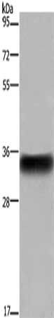
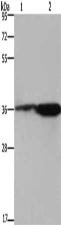
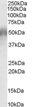
| Application notes | ELISA: Peptide ELISA: antibody detection limit dilution 1:16000.WB: Approx 34kDa band observed in Human myeloid erythroblast cell line K562 and Human monocytic cell line U937 lysates (calculated MW of 34.0kDa according to NP_003992.3;). In transfected HEK293 transiently expressing CD32 a band of approx. 40kDa (glycoprotein) is observed. This band is not observed in the non-transfected HEK293. Recommended concentration: 0.3-1 μg/ml. |
| Expiration Date | 12 months from date of receipt. |
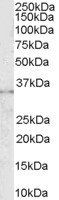
Western blot analysis of K562 lysate using CD32b antibody
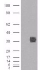
Western blot analysis of HEK293 lysate using CD32b antibody
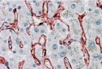
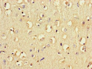
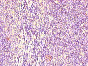
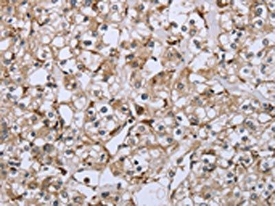
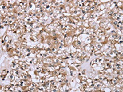
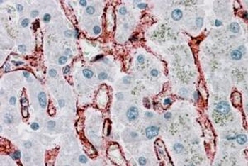
Immunohistochemical staining of Human Liver using CD32b antibody
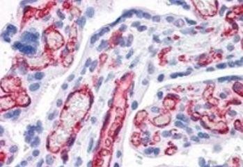
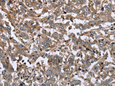
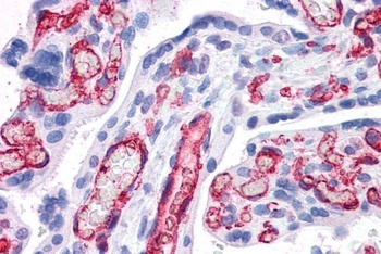
Immunohistochemical staining of Human Placenta using CD32b antibody
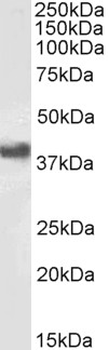
orb19010 (1 µg/mL) staining of Daudi cell lysate (RIPA buffer, (35 µg protein in RIPA buffer). Detected by chemiluminescence.
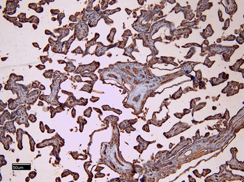
orb19010 (8 µg/mL) staining of paraffin embedded Human Placenta. Heat induced antigen retrieval with citrate buffer pH 6, HRP-staining.
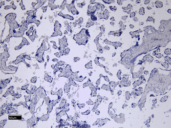
orb19010 Negative Control showing staining of paraffin embedded Human Placenta, with no primary antibody.
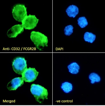
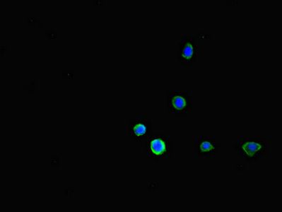
orb19010 Immunofluorescence analysis of paraformaldehyde fixed THP-1 cells immobilized on ShifixTM coverslip, permeabilized with 0.15% Triton. Primary incubation 1 hr (10 µg/mL) followed by Alexa Fluor 488 secondary antibody (2 µg/mL), showing membrane staining. The nuclear stain is DAPI (blue). Negative control: Unimmunized goat IgG (10 µg/mL) followed by Alexa Fluor 488 secondary antibody (2 µg/mL).
- Item 1 of 7
CD32b antibody [orb14369]
ICC, IF, IHC-P, WB
Guinea pig, Mouse, Rat
Rabbit
Polyclonal
Unconjugated
100 μg, 200 μg - Item 1 of 3
- Item 1 of 3
- Item 1 of 3
- Item 1 of 3


Submit a review
Filter by Rating

- 5 stars

- 4 stars
- 3 stars
- 2 stars
- 1 stars
